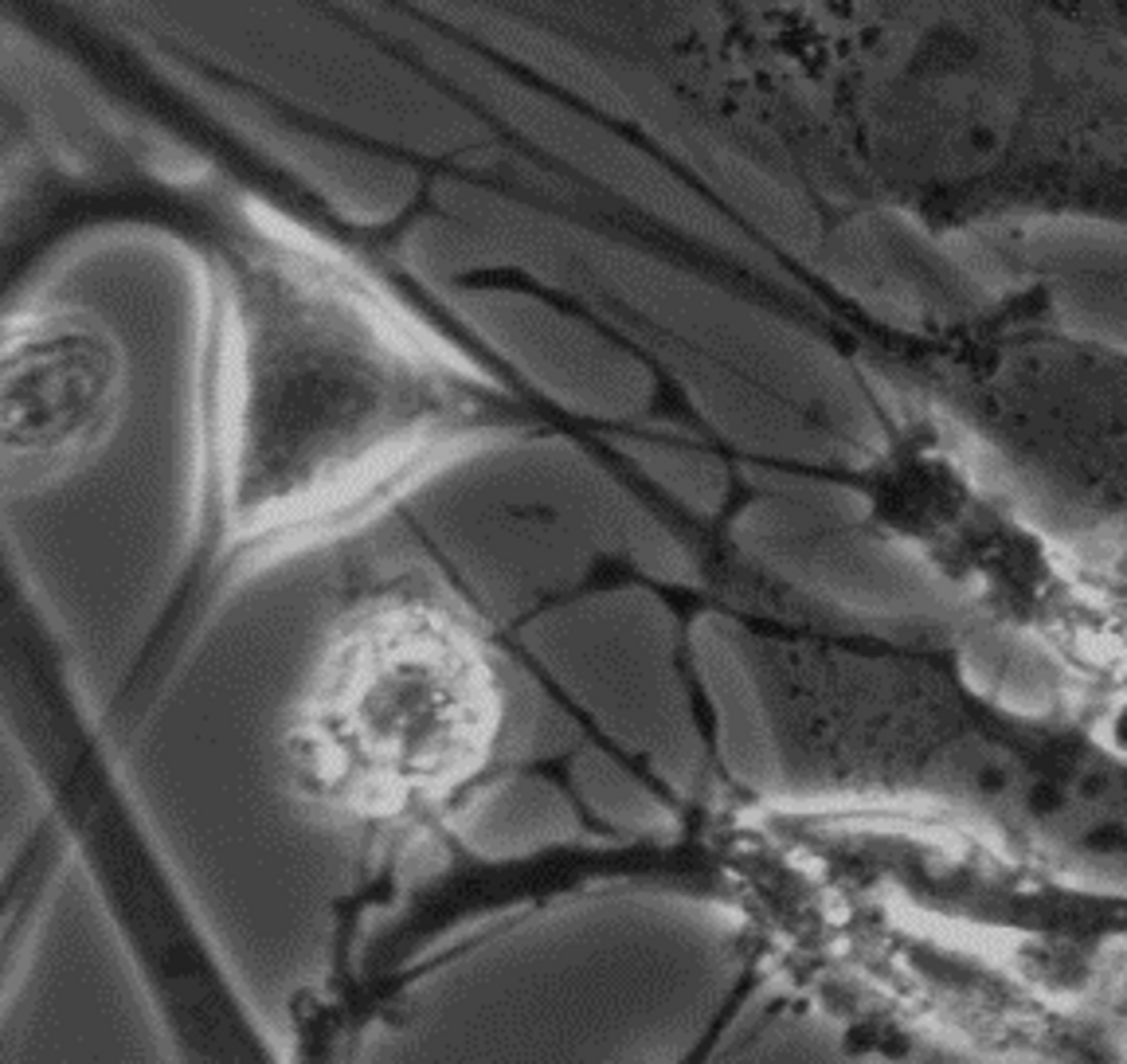
The myenteric plexus is a mesh-like network of nerve cells embedded between the muscle layers of your digestive tract. It acts as the primary motor controller for gut movement, coordinating the muscular squeezes that drive peristalsis. While it can run digestion entirely on its own, it also communicates directly with the central nervous system.
By the edgi team We find the most surprising true thing about an idea and build a 60-second lesson around it.
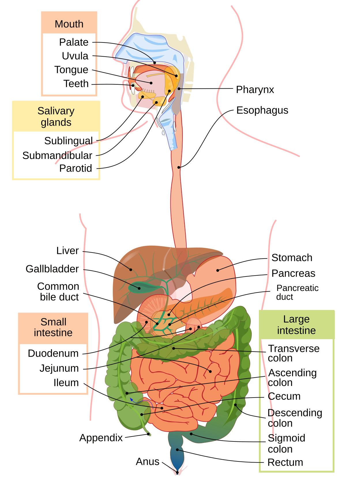
Stretching from your esophagus to your anus is a massive, independent network of about 500 million neurons. It contains more nerve cells than your spinal cord and can sense, integrate, and execute actions without any input from your actual head.
A diagram of the human digestive system, labeling organs from the mouth to the anus. Mariana Ruiz, Jmarchn, Public domain, via Wikimedia Commons
Embedded deep within the muscular walls of your digestive tract is a dense mesh of nerve fibers called the myenteric plexus. First discovered by the German neuropathologist Leopold Auerbach, it acts as the primary motor controller for gut movement.
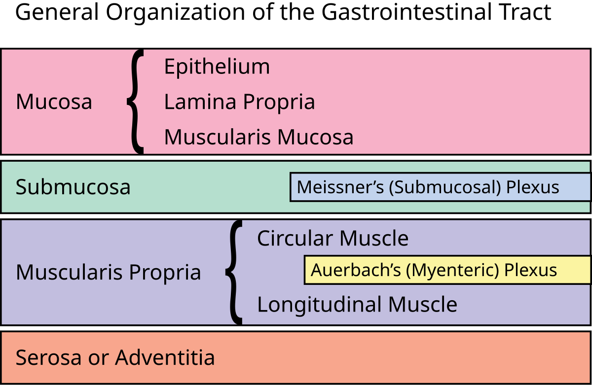
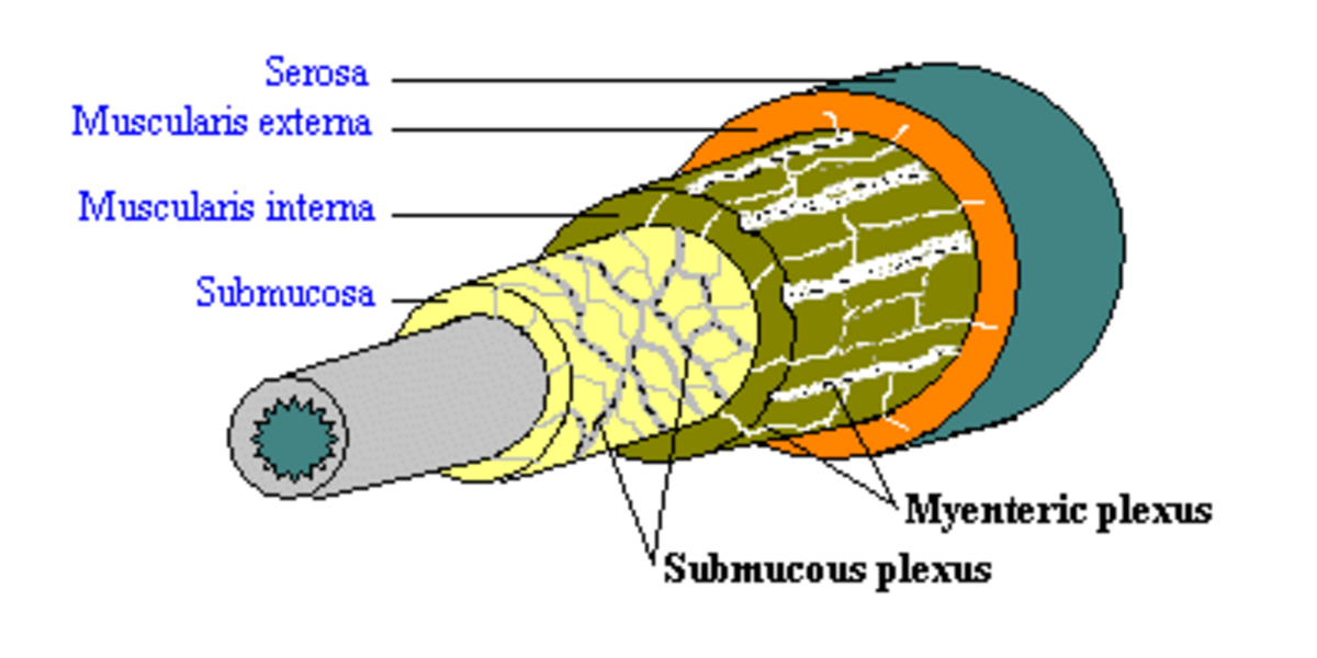
Diagram titled "General Organization of the Gastrointestinal Tract" showing the hierarchical layers of the GI tract. Rehua, CC BY-SA 3.0, via Wikimedia Commons
It coordinates the rhythmic squeezing and relaxing required to push food forward through a process called peristalsis.
How it works
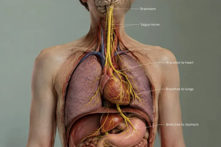
The myenteric plexus receives signals from both sympathetic and parasympathetic pathways, tuning gut activity to your broader physical state. Right next to it sits the submucosal plexus, which manages local secretions and blood flow rather than raw muscle movement.
An anatomical illustration depicting the submucosal plexus, showing a network of nerve cells and fibers. Henry Vandyke Carter, Public domain, via Wikimedia Commons
Together, these layered nerve networks ensure digestion runs smoothly even when your central nervous system is completely distracted.
Where the myenteric plexus sits and what it contains
The myenteric plexus sits between the circular and longitudinal muscle layers of the muscularis externa throughout the esophagus, stomach, and intestine. Like the central nervous system, its ganglia contain glia, interneurons, a dense synaptic neuropil, and Dogiel cells, all kept isolated from blood vessels.
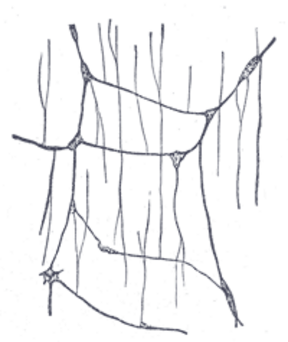
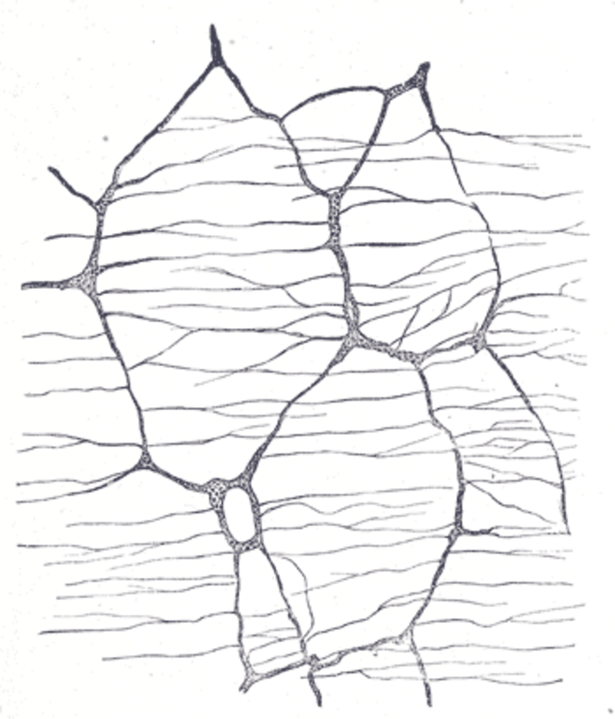
A mesh-like network of interconnected ganglia and nerve fibers forms the myenteric plexus within the muscular wall. Henry Vandyke Carter, Public domain, via Wikimedia Commons
Around 30% of its neurons are enteric sensory neurons, meaning it senses local gut states in addition to issuing motor commands. Special pacemaker cells called interstitial cells of Cajal generate slow waves of basal electrical rhythm, controlling the timing of peristaltic and segmental muscle contractions.
How it controls digestion and communicates
The enteric nervous system makes use of more than 30 neurotransmitters, including acetylcholine, dopamine, and serotonin. In fact, over 90% of the body's serotonin and roughly 50% of its dopamine reside in the gut. All myenteric neurons express nicotinic cholinergic receptors, while specific subsets express other targets like the 5-HT4 serotonin receptor, which can be activated to stimulate motility.
The myenteric plexus rests between the muscle layers, operating alongside the nearby submucous plexus. R. Bowen, Public domain, via Wikimedia Commons
The system also carries taste receptors like TAS1R3 and the taste protein gustducin. While these sense sweetness on the tongue, in the gut they sense glucose and help regulate insulin secretion. External cues feed into this network as well: seeing appealing food prompts the central nervous system to trigger stomach secretions through shared nerve links.
What happens when myenteric neurons fail
Damage to the myenteric plexus leads to severe motility disorders. In Hirschsprung's disease, neuroblasts fail to migrate during development, leaving a section of the lower bowel without myenteric or submucosal ganglion cells and creating a bowel obstruction. In achalasia, a drop in ganglion cell density within the esophageal myenteric plexus disrupts normal swallowing.
Because of the close parallels between the enteric and central nervous systems, gut tissue provides a window into broader neurological conditions. People with Parkinson's disease often experience severe constipation years before motor symptoms appear, leading researchers to examine colonic biopsies to study the condition.
Test yourself
Does the brain in your head directly control every single gut movement?
No, it operates independently. Your gut contains a massive, independent nerve network with 500 million neurons that can manage digestion without any input from your head.
Why might an animal digest food successfully even when deeply unconscious?
Local neural circuits drive motion. Autonomic local neural networks ensure essential physiological processes run smoothly even when the central nervous system is entirely distracted.
Does your gut need signals from your head to push food along?
No, it manages movement alone. The enteric nervous system handles the entire rhythmic process of digestion locally, without needing instructions from the brain in your head.
Play the lesson in edgi and the card is yours. It lands on your Map next to the ideas it connects to, and turns from matte to foil to gold as you learn more around it.
What is the difference between the myenteric plexus and the submucosal plexus?
The myenteric plexus sits between the outer muscle layers and primarily controls muscular contractions and motility. The submucosal plexus sits closer to the inner lining of the gut, where it regulates local secretions and blood flow.
Who discovered the myenteric plexus?
The German neuropathologist Leopold Auerbach first discovered it using histological staining techniques. Because of his work, it is also known as Auerbach's plexus.
Where do myenteric neurons come from during development?
Myenteric neurons originate from the vagal neural crest during embryonic development. From there, developing nerve cells migrate down the digestive tract to form the network.