REM sleep is a distinct phase of sleep where the brain acts nearly awake while the body's skeletal muscles are paralyzed. It is also known as paradoxical sleep because its fast electrical brainwaves look like wakefulness rather than deep rest. During this state, core body temperature rises, heart rate turns irregular, and the most vivid dreams occur.
By the edgi team We find the most surprising true thing about an idea and build a 60-second lesson around it.
In 1953 Eugene Aserinsky and Nathaniel Kleitman reported something that had been in front of everyone forever. Sleepers' eyes move, in bursts, several times a night. The movements were quick and jerky, not the slow drift of someone falling asleep. Aserinsky had first caught them while testing his recording equipment on his own young son.
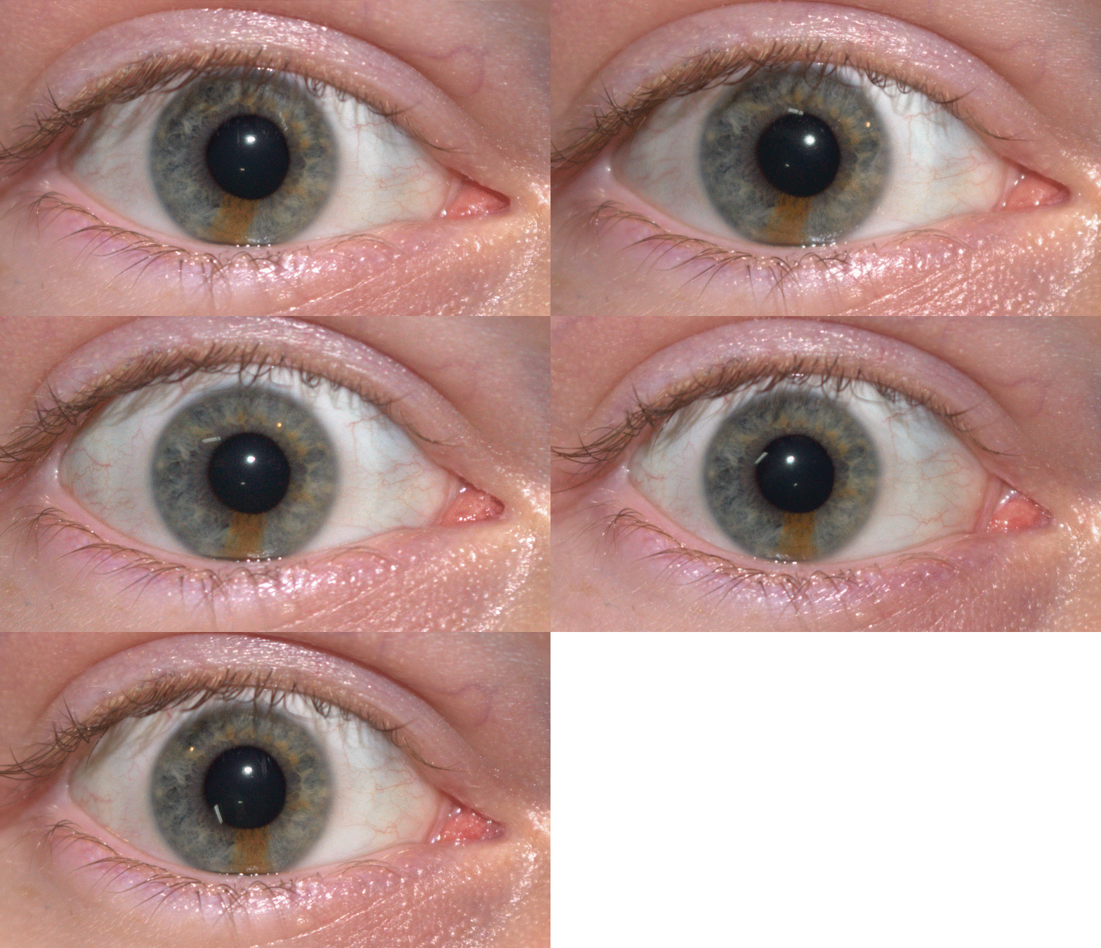
A montage of five pictures of a male subject's right eye demonstrating torsional eye movement, with partial heterochromia visible in the iris. Dingolover6969, CC0, via Wikimedia Commons
During those bursts the brain's electrical trace, the EEG, looked closer to waking than to deep sleep, and breathing and heart rate turned irregular. They named the state after the eyes: REM sleep.
The body is switched off
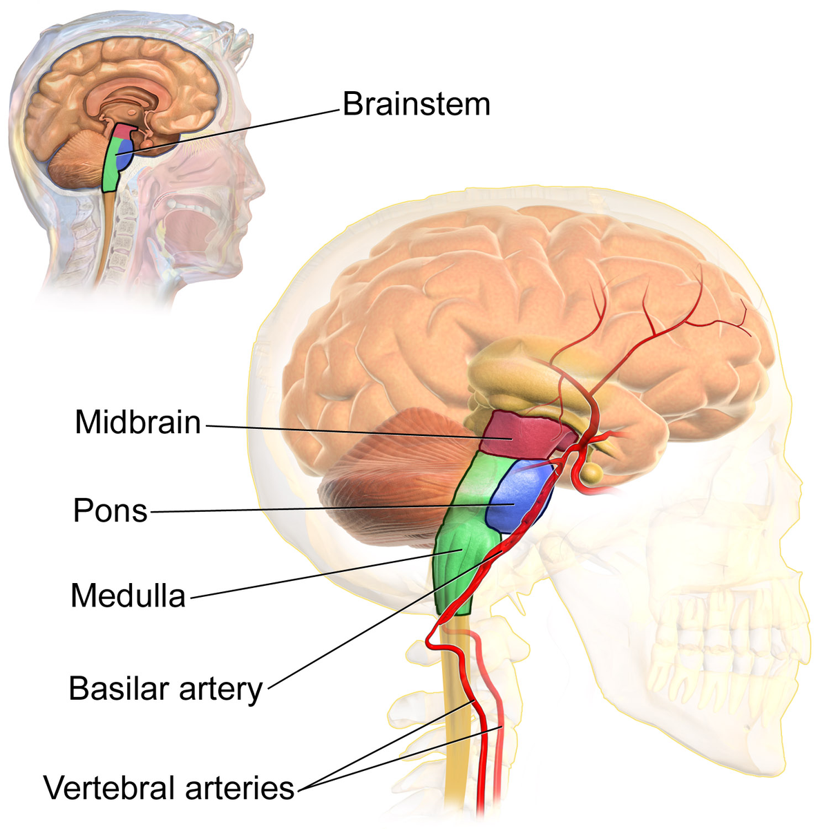
While that is going on, the muscles that hold you up go slack. Tone in the arms, legs and neck falls to almost nothing for the length of the bout. Nothing is disconnected. Cells in the brainstem send an active inhibiting signal down onto the motor neurons, which is a hand held on the switch rather than a wire pulled out.
A diagram illustrates the anatomy of the human brainstem and its associated arteries, with an inset showing its position within the skull. BruceBlaus. When using this image in external sources it can be cited as: Blausen.com staff (2014). "Medical gallery of , CC BY 3.0, via Wikimedia Commons
The muscles you cannot spare are exempt. The diaphragm keeps breathing you, the heart is not skeletal muscle at all, and the small muscles moving the eyes are left free, which is the part you can see.
When the switch fails
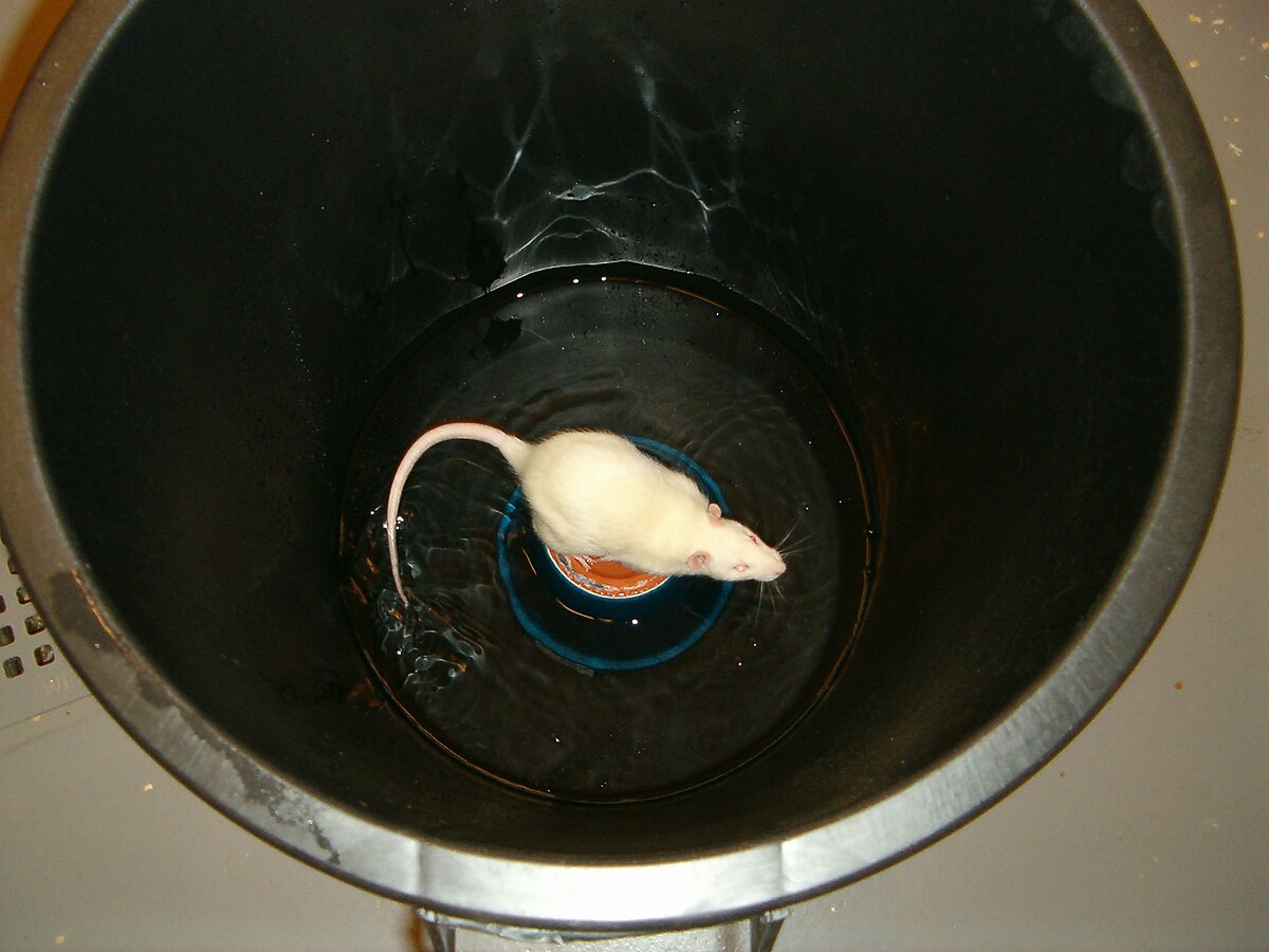
In rapid eye movement sleep behavior disorder the paralysis does not arrive. People kick, shout, punch and climb out of bed while still asleep, and what they do matches what they report on waking. The same state can be produced on purpose: silence those inhibitory brainstem cells in a rat and it enters REM sleep with its muscles working. The stillness is something the brain does.
A white laboratory rat is shown in a "flowerpot technique" setup, standing on a small platform in a bucket of water, a method used to deprive rats of REM sleep. Jean-Etienne Poirrier, CC BY-SA 2.5, via Wikimedia Commons
Where it sits in a night
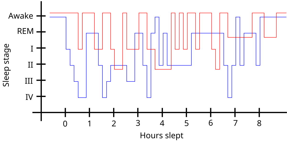
REM arrives in bouts. The first is roughly 90 minutes after you fall asleep and runs about ten minutes; each one after it is longer, and the longest come towards morning. So an alarm that cuts the night short takes mostly this stage. The deepest sleep, the slow-wave sleep of the first hours, has already happened.
Graph comparing sleep patterns, with Sleep stage on the Y-axis (Awake, REM, I, II, III, IV) and Hours slept on the X-axis (0 to 8.5 hours). RLS-Schlafmuster.png: Markus Mueller derivative work: -- Editor at Large • talk, CC BY-SA 3.0, via Wikimedia Commons
The most vivid, story-shaped dreams get reported out of REM. People woken from non-rapid eye movement sleep report dreams too, often about half the time, so REM is not the only room this happens in.
Why is it called paradoxical sleep?
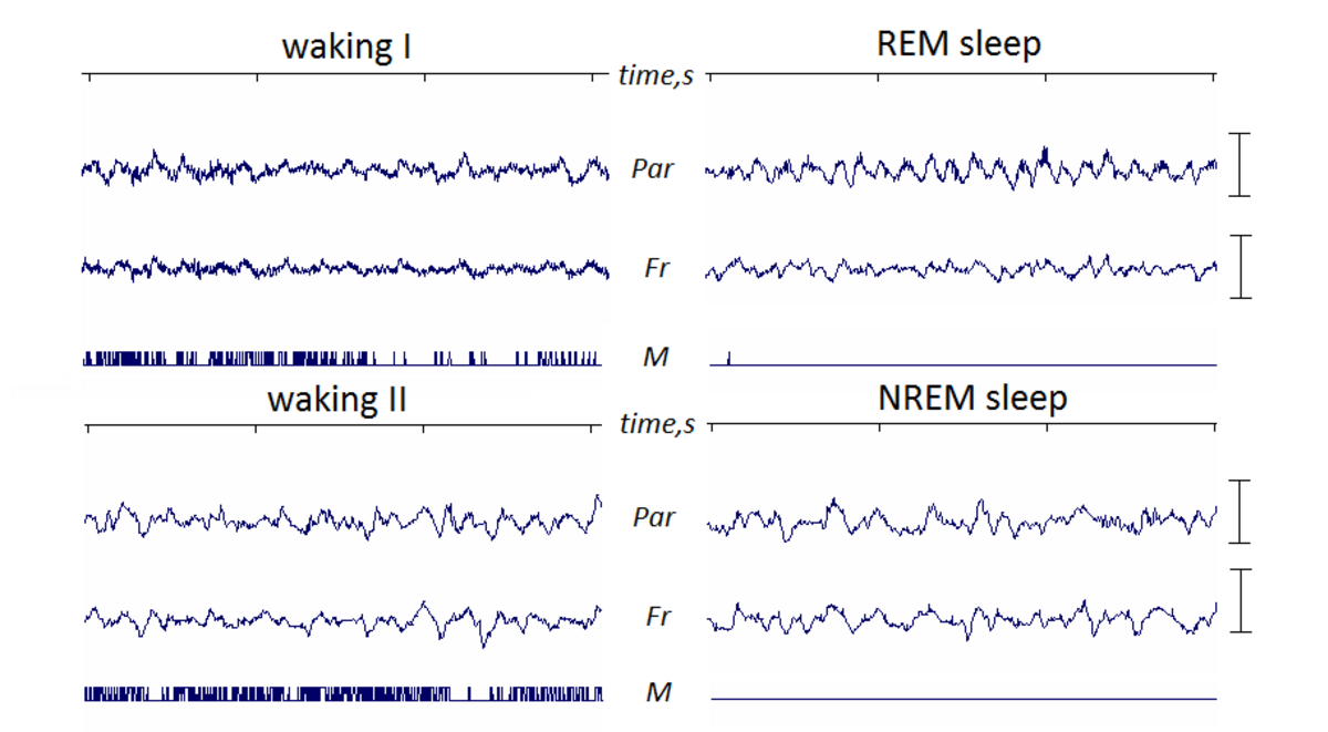
Brain energy consumption during REM sleep equals or exceeds energy use in wakefulness, burning 11 to 40 percent more glucose and oxygen than slow-wave sleep. Instead of the slow delta waves of deep non-REM sleep, an electroencephalogram (EEG) shows fast, low-voltage, desynchronized waves. Prominent 3 to 10 Hz theta rhythms dominate the hippocampus and cortex, alongside fast 40 to 60 Hz gamma waves.
An EEG recording of a mouse during REM sleep shows the prominent, fast theta rhythm that distinguishes this stage from deep sleep. Andrii Cherninskyi, CC BY-SA 4.0, via Wikimedia Commons
Neurons in the cortex and thalamus are depolarized, meaning they fire more readily than during deep sleep. At the same time, frontal and posterior areas show looser electrical connectivity across most frequencies, mirroring the disorganized flow of dreams. Posterior regions and both brain hemispheres remain highly coherent with each other, especially during lucid dreaming.
What chemicals and brain areas drive REM sleep?
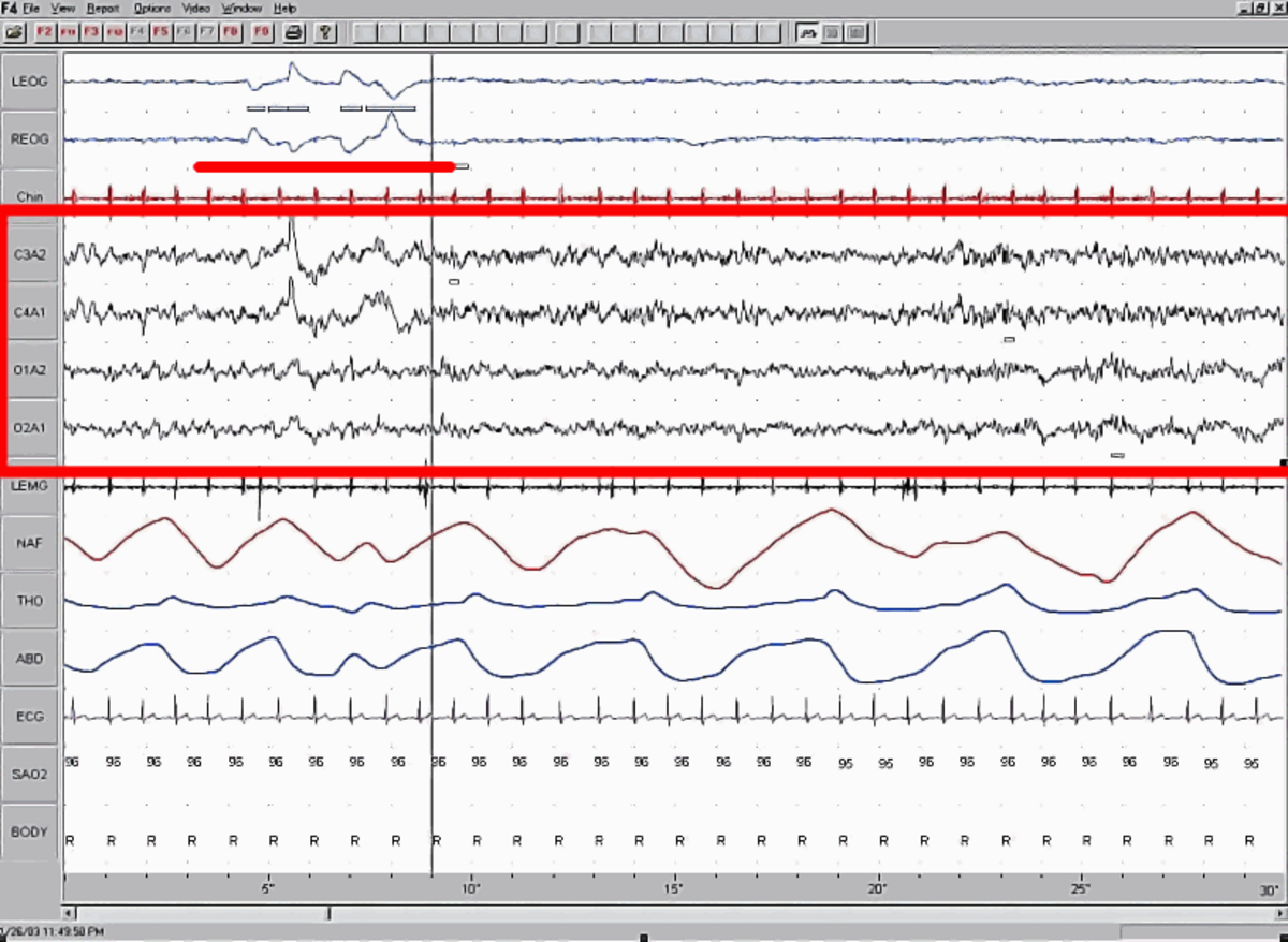
The shift into REM sleep starts in the brainstem, particularly the pontine tegmentum and locus coeruleus. Bursts of electrical spikes called ponto-geniculo-occipital (PGO) waves originate there in clusters every six seconds during the transition into paradoxical sleep. These waves peak in amplitude as they travel to the visual cortex, driving the rapid bursts of eye movement.
A polysomnogram traces rapid eye movements along the red line while recording fast, low-voltage brain activity inside the red box. MrSandman at English Wikipedia, Public domain, via Wikimedia Commons
This state runs on a unique chemical mixture. The brainstem floods the system with acetylcholine, the chemical messenger linked to rapid brainwaves, while nearly shutting off monoamines like serotonin, histamine, and norepinephrine. Because norepinephrine is almost completely absent, dreams and experiences from REM sleep are not transferred into permanent memory.
Inside the forebrain, PET scans reveal high activity in limbic and paralimbic structures tied to memory, emotion, fear, and sex. The amygdala stays active, helping generate PGO waves and regulating cardiac rhythms, while areas involved in complex thought show activity levels matched only in waking hours.
How does REM sleep change the body?
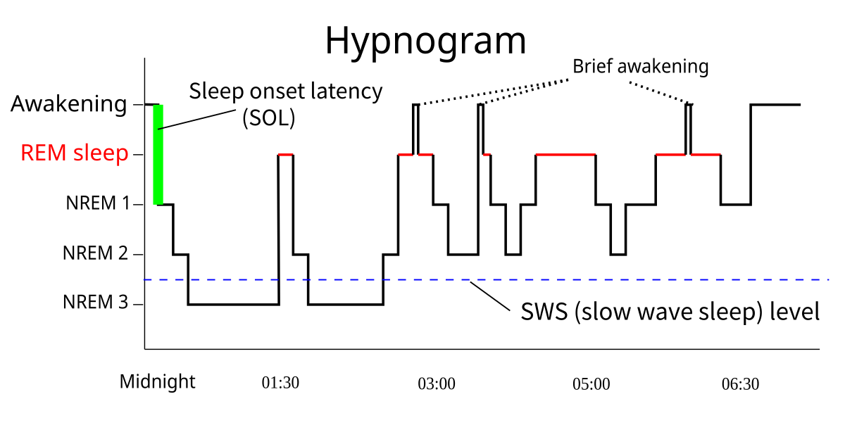
Organisms in REM sleep suspend central homeostasis. Breathing turns irregular, skin temperature drops to its lowest values, and core body and brain temperatures rise. In a typical seven-hour sleep period, REM sleep occurs four times, taking up an increasing fraction of each 90-minute cycle as morning approaches.
A hypnogram maps how alternating 90-minute sleep cycles shift toward longer proportions of paradoxical REM sleep later in the night. RazerM, CC BY-SA 3.0, via Wikimedia Commons
Test yourself
During REM sleep, is muscle relaxation caused by severed motor pathways?
False. Motor pathways are not severed or disconnected. Brainstem cells actively inhibit motor neurons, holding the switch down while the brain remains highly active.
What characterizes electrical and physiological activity during REM sleep?
Brain activity resembles waking states. During REM sleep, brain electrical traces resemble waking patterns while breathing and heart rate turn irregular rather than steady.
What happens to the postural muscles during REM sleep?
The brainstem actively holds them down. The atonia is produced by inhibitory brainstem cells signalling the motor neurons. Nothing is unplugged.
Play the lesson in edgi and the card is yours. It lands on your Map next to the ideas it connects to, and turns from matte to foil to gold as you learn more around it.
What happens if someone is repeatedly woken during REM sleep?
Waking a person every time they enter REM sleep produces REM deprivation. When permitted to sleep without interruption again, test subjects experience a modest REM rebound, spending more time in the stage than normal.
Do other animals experience REM sleep?
All mammals and birds experience REM sleep. It is also found in some reptiles.
Why do dreams fade so quickly upon waking?
The neurotransmitter norepinephrine is almost completely absent from the brain during REM sleep. Without this chemical, the brain cannot transfer dream experiences into permanent memory storage.