AutophagyHow cells survive by eating their own parts
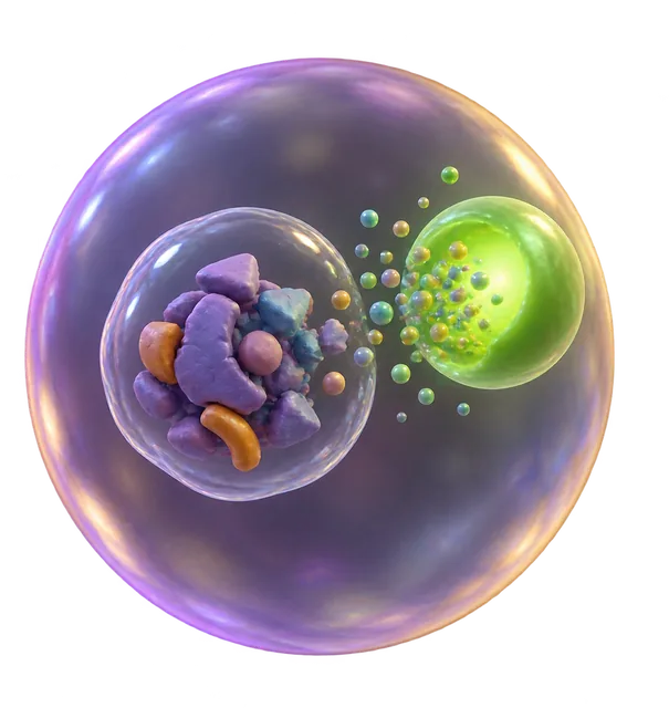
Autophagy is the process biological cells use to clean house by breaking down and recycling their own damaged or unnecessary parts. A cell isolates cellular trash inside a specialized bubble and ships it to an organelle called a lysosome, which breaks the debris down into raw building blocks. Beyond providing survival fuel during starvation, this recycling runs continuously to clear out toxic clutter that contributes to disease and aging.
By the edgi team We find the most surprising true thing about an idea and build a 60-second lesson around it.
In the 1990s, Yoshinori Ohsumi spent years staring at baker's yeast under a microscope to solve a mystery: where does cellular waste go? He engineered yeast that couldn't break down its trash, then starved the cells to trigger their survival instincts.
Portrait of Yoshinori Ohsumi, a Nobel Prize laureate, shown from the chest up, wearing glasses and a grey jacket, with a microphone near his right ear. Bengt Nyman from Vaxholm, Sweden, CC BY 2.0, via Wikimedia Commons
He watched as tiny sacs appeared, swelling with the cell's own parts as they headed for the lysosome to be destroyed. This discovery, that cells have a genetically programmed self-eating system, eventually earned him a Nobel Prize.
The recycling plant
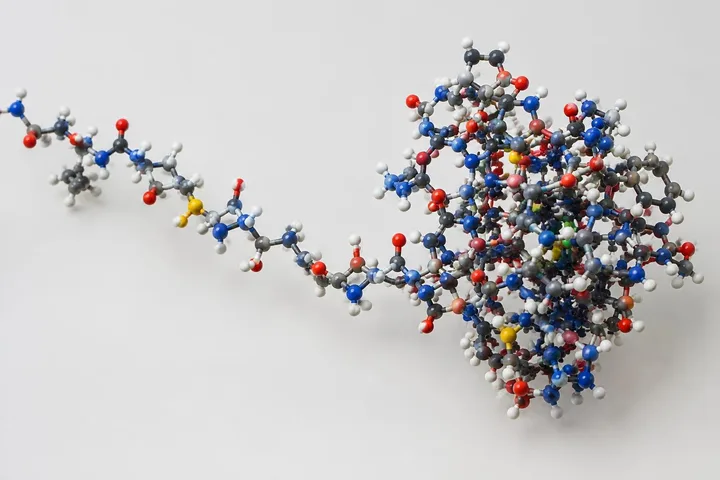
The process is called autophagy, which literally means self-devouring. It is not just for starvation; it is constant house-cleaning. Cells form a double-membrane bubble around trash, like broken proteins, and ship it to the lysosome.
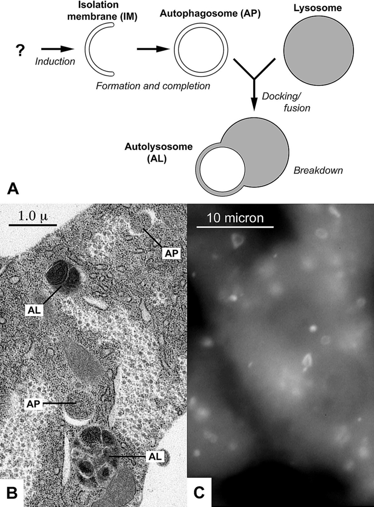
A multi-panel image illustrates the process of autophagy, showing a diagram (A), an electron micrograph (B), and a fluorescently labeled image (C). Juhasz G, Neufeld TP. Original images by Ryan Scott (B) and Dr. Noboru Mizushima (C)., CC BY 2.5, via Wikimedia Commons
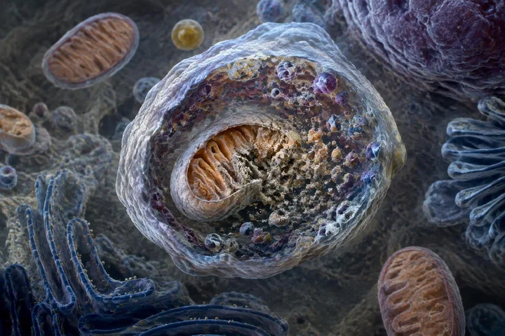
The lysosome acts like a molecular trash compactor, breaking the debris into raw building blocks. By recycling this cellular junk, the body prevents the toxic buildup that eventually leads to aging and disease.
The master switch
A protein called mTOR acts as the master switch for this cleaning crew. When you eat, mTOR signals the cell to grow and build, effectively pausing the sweepers.
When you practice fasting, mTOR levels drop, and the cellular sweepers go into overdrive, pruning away the damaged parts that cause cellular senescence.
A glass of water sits on a white, empty plate, flanked by a fork on the left and a knife on the right, all placed on a wooden table outdoors. Dr Jean Fortunet, CC BY-SA 3.0, via Wikimedia Commons
How does autophagy work inside a cell?
In macroautophagy, the primary form of this process, the cell isolates its target using a curved structure called a phagophore. This structure wraps around cytoplasmic debris, such as worn-out mitochondria or damaged proteins, sealing them within a double-membrane bubble called an autophagosome.
The autophagosome then travels to and fuses with an available lysosome, forming a hybrid structure called an autolysosome. Inside, the lysosome delivers specialized enzymes that break the trapped contents down into basic molecular components for the cell to reuse.
The entire system is regulated by nutrient availability through master switch proteins like mTOR. When nutrients are plentiful, mTOR promotes growth and limits recycling, but when food drops or stress occurs, autophagy accelerates to protect cellular energy levels.
What are the different types of autophagy?
Scientists categorize autophagy into four distinct forms: macroautophagy, microautophagy, chaperone-mediated autophagy (CMA), and crinophagy. Macroautophagy is the most extensively studied pathway, while crinophagy specializes in degrading unnecessary secretory granules.
Macroautophagy itself divides into bulk and selective varieties. While bulk autophagy engulfs general cytoplasmic material during starvation, selective autophagy targets specific cellular components for destruction, such as mitophagy for mitochondria, lipophagy for fats, and pexophagy for peroxisomes.
Why is autophagy linked to human disease?
Defects in autophagy are tied directly to conditions like neurodegeneration and cancer. In 1999, Beth Levine's research team published a landmark discovery formally connecting autophagy genes to cancer development.
Autophagy plays a dual role in human health. It can act as an adaptive survival response that protects stressed cells from toxic buildup, but under certain conditions, it can also promote cell death and morbidity.
Test yourself
When a complex biological system faces starvation, why might it actively destroy its own parts?
To harvest raw materials for vital survival. Cells do not just shut down or die; they selectively consume damaged internal components to harvest raw blocks for essential functions.
Starving a cell causes it to consume its own functional parts to die.
False. Cells do not eat themselves to die; they perform a strategic recycling process to survive, stripping away only the damaged components to create fresh resources.
Play the lesson in edgi and the card is yours. It lands on your Map next to the ideas it connects to, and turns from matte to foil to gold as you learn more around it.
Belgian biochemist Christian de Duve coined the term in 1963 after discovering the functions of lysosomes. He established that lysosomes are the sites responsible for glucagon-induced cellular degradation in the liver.
What genes control autophagy?
Autophagy is controlled by a set of autophagy-related genes, officially standardized under the unified name ATG in 2003. These genes were first mapped out in budding yeast during the 1990s by researchers including Yoshinori Ohsumi.