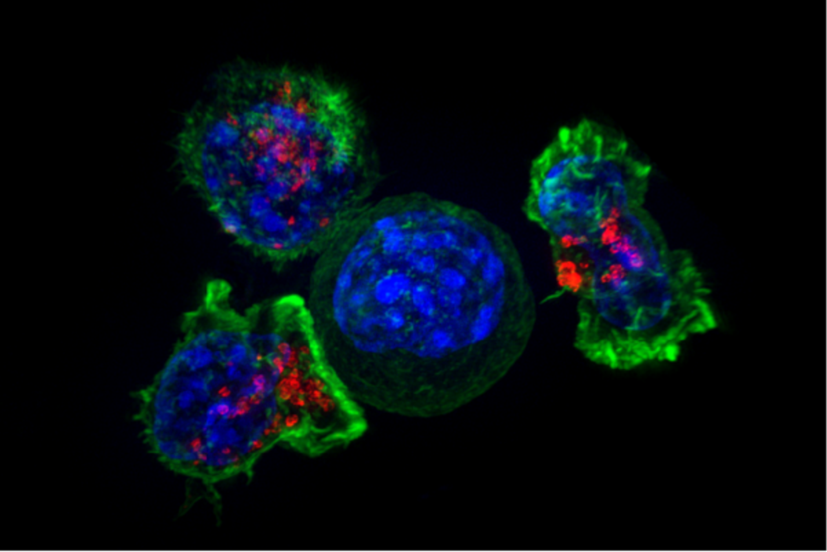
The sculptor
When you were a developing embryo, your hands looked like solid paddles. To separate your fingers, the cells in the webbing had to disappear.
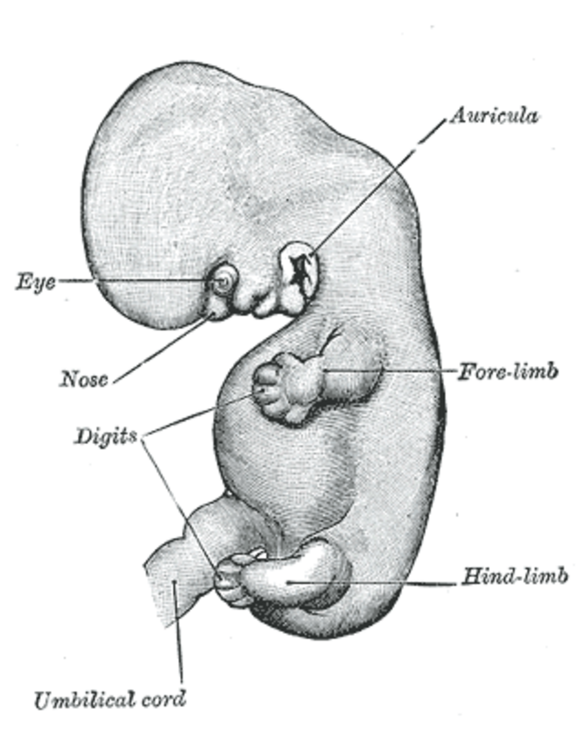
They didn't just die by accident; they were given a genetic order to commit suicide.
Apoptosis is programmed cell death, a built-in biological routine where an unneeded or damaged cell systematically destroys itself. Instead of bursting and harming its neighbors, the dying cell neatly shrinks, breaks apart, and signals scavenger cells to consume the remains. An average adult quietly eliminates between 50 and 70 billion cells this way every single day.

By the edgi team We find the most surprising true thing about an idea and build a 60-second lesson around it.
When you were a developing embryo, your hands looked like solid paddles. To separate your fingers, the cells in the webbing had to disappear.
They didn't just die by accident; they were given a genetic order to commit suicide.
When cells die from injury, they burst in a process called necrosis, spilling toxic waste and causing inflammation. Apoptosis is different. It is controlled, orderly, and clean.
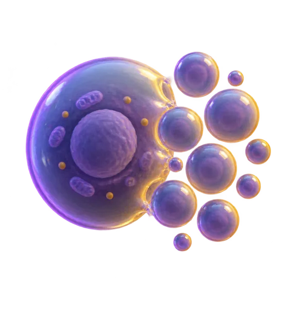
The cell shrinks, packages its contents into neat bundles, and signals neighbors to eat it.
Scientists first realized this was a program, not a malfunction, by studying a transparent worm called C. elegans. They counted exactly 1,090 cells in the worm's body.
They discovered that exactly 131 of those cells were destined to die as part of normal development.
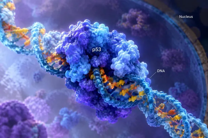
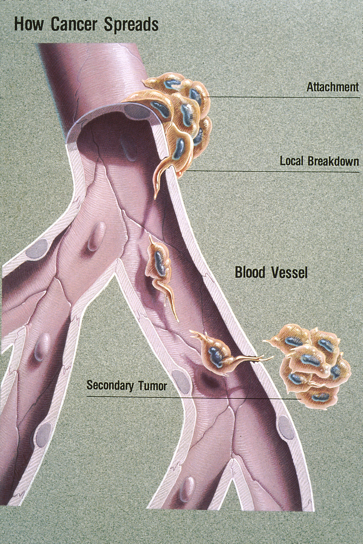
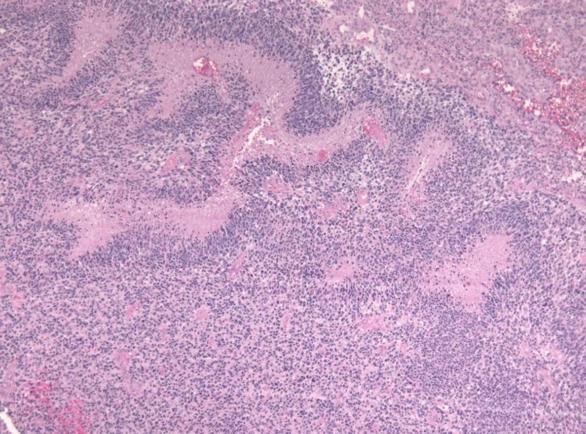
This self-destruct switch is a critical safety net. When it breaks, often due to an oncogene, damaged cells refuse to die and begin to multiply uncontrollably.
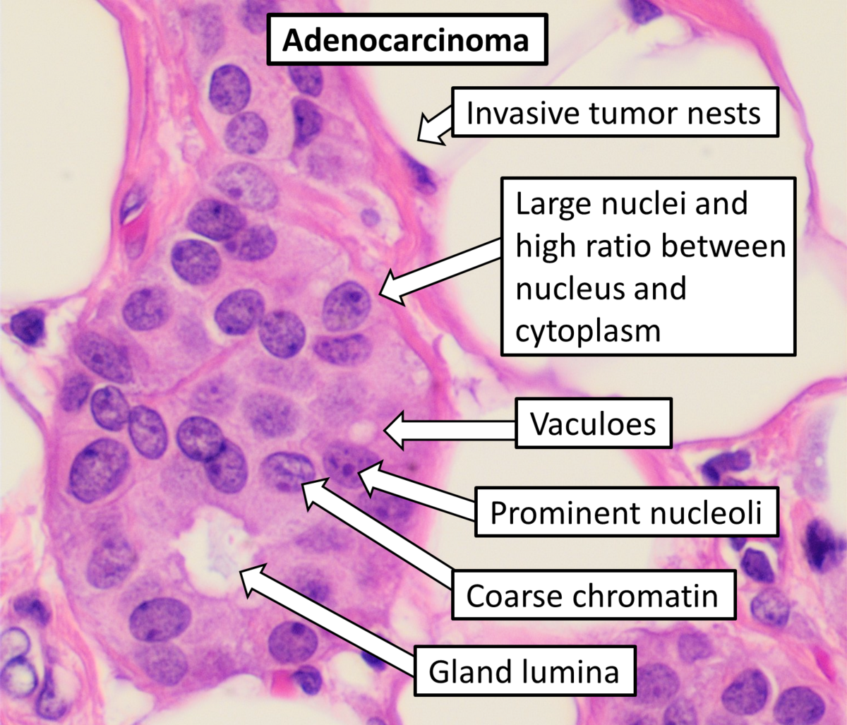
Cancer is, in many ways, the failure of this vital suicide command.
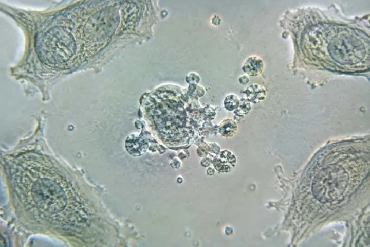
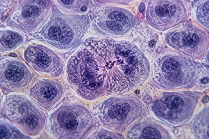
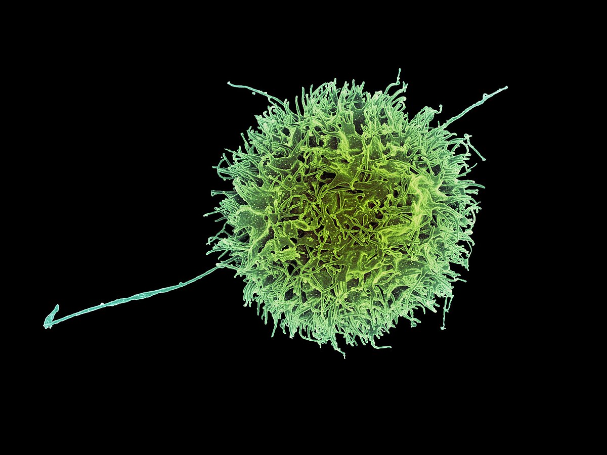
A dying cell does not simply break open. During apoptosis, a cell shrinks, its DNA and chromatin condense and fragment, its messenger RNA decays, and its outer membrane forms irregular bulges called blebs.
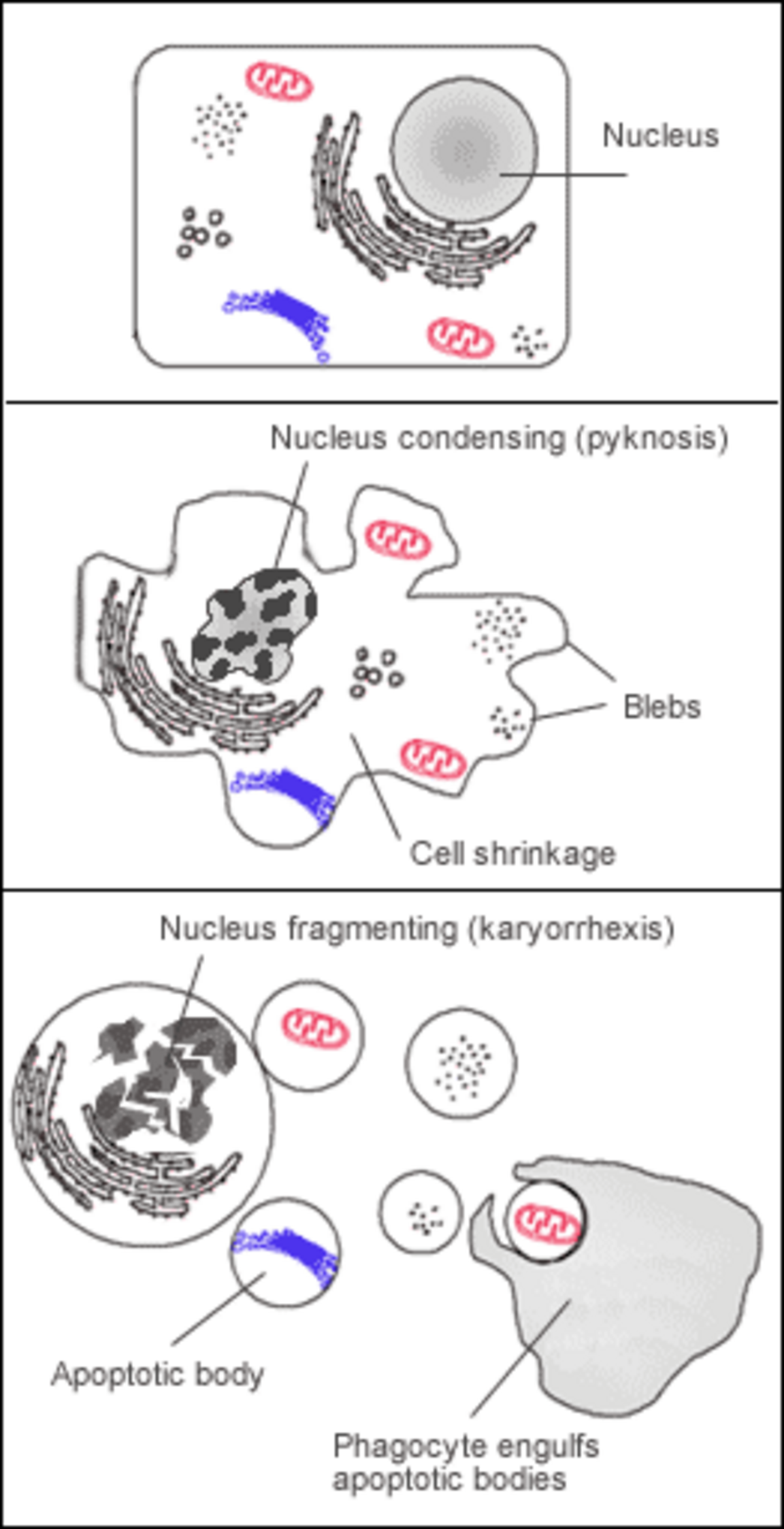
The cell then packages its parts into neat membrane-bound fragments known as apoptotic bodies. Specialized scavenger cells called phagocytes engulf and clear these fragments before toxic internal contents can spill into the surrounding tissue. This orderly cleanup prevents the inflammation and tissue damage caused by necrosis, which is accidental cell death from physical trauma or injury.
Because apoptosis cannot stop once it starts, cells keep its trigger under strict control. The process begins through either an intrinsic pathway, where the cell senses internal stress and releases proteins from its mitochondria, or an extrinsic pathway, where signals from neighboring cells bind to cell-surface death receptors.
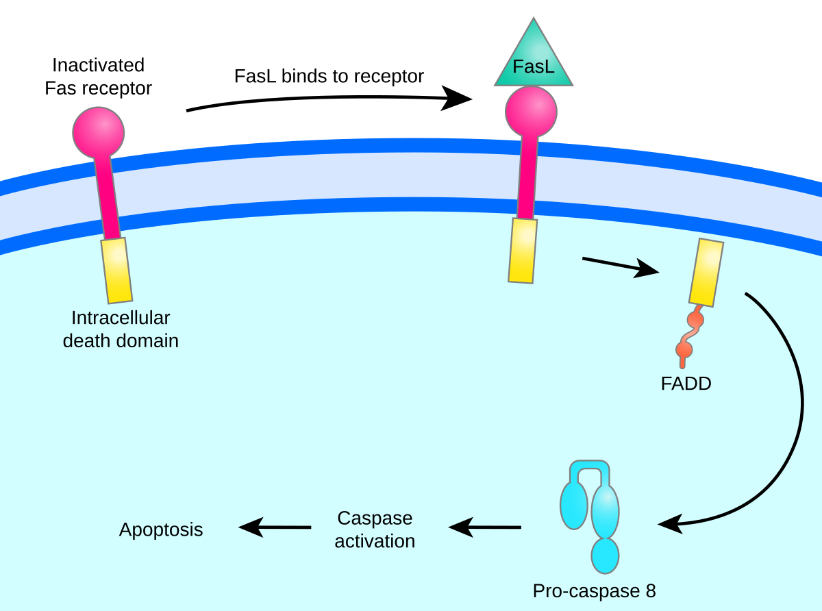
Both pathways activate caspases, which are specialized enzymes that break down proteins. Initiator caspases switch on first, which in turn activate executioner caspases. These executioners tear through cellular proteins indiscriminately, ensuring complete destruction.
Cell survival depends on a precise balance between death-promoting factors like caspases and Fas receptors, and death-blocking proteins from the Bcl-2 family. Tilting this balance in either direction causes severe disease.
When apoptosis is hyperactive, excessive cell loss causes organ and tissue atrophy. When apoptosis fails, damaged cells refuse to self-destruct and multiply unchecked. This breakdown is a hallmark of cancers like follicular lymphoma, where the BCL2 gene produces excess protein that blocks the cell suicide command.
In apoptosis, how does the cell process differ from accidental injury?
It packages its contents neatly for neighbors.. Apoptosis is a controlled process where the dying cell packages itself neatly, preventing toxic waste spills and inflammation.
Is apoptosis defined by the messy release of cell contents into the body?
No, it is a clean process.. Apoptosis is an orderly, controlled process where cells package themselves for removal; necrosis is the messy process involving cell bursting and inflammation.
The elimination of the webbing between human fingers during embryonic development is a classic example of ___.
apoptosis. Sculpting our hands and feet requires the precise removal of cells that are no longer needed.
Collectible card
Play the lesson in edgi and the card is yours. It lands on your Map next to the ideas it connects to, and turns from matte to foil to gold as you learn more around it.
The word comes from an Ancient Greek term meaning 'falling off', originally used to describe leaves falling from trees or petals dropping from flowers. Greek language professor James Cormack suggested the name to researchers in 1972 to describe natural, orderly cell death.
An average human child between 8 and 14 years old loses approximately 20 to 30 billion cells to apoptosis every day. Multicellular organisms use this constant turnover for healthy growth and tissue remodeling.
Sydney Brenner, H. Robert Horvitz, and John Sulston won the 2002 Nobel Prize in Medicine for identifying the genes that control apoptosis in the transparent nematode worm C. elegans. Humans possess equivalent versions of these same regulatory genes.
Part of the Set · 8 cards
A tumor isn't an invader. It's your own cells, mutating and competing by the exact rules that built every species on Earth.
Part of the Set · 8 cards
Almost every cell in your body carries an ID badge. Show the wrong one, or none, and your own immune system kills it.
Learn the whole SetLesson complete
You finished the lesson. In edgi, the card goes on your Map next to everything else you learn.