Cell CultureGrowing living tissues outside the body
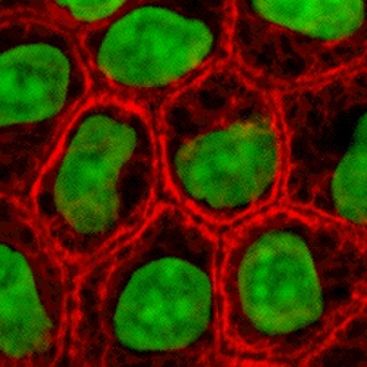
Cell culture is the process of growing living cells outside their natural organism inside controlled laboratory vessels. Scientists isolate these cells from animal or plant tissues and feed them precise mixtures of nutrients, gases, and warmth at body temperature. This technique allows researchers to study diseases, test new drugs, and produce vaccines without experimenting directly on living patients.
By the edgi team We find the most surprising true thing about an idea and build a 60-second lesson around it.
In 1951, a researcher named George Gey received a sample of tumor cells from a patient named Henrietta Lacks. Before this, human cells in a lab died within days. But these cells, now called HeLa, kept dividing indefinitely.
A black and white portrait of Henrietta Lacks, showing her smiling slightly and wearing a dark jacket with a light-colored collar. Oregon State University, CC BY-SA 2.0, via Wikimedia Commons
They were the first immortalized cell line, a biological breakthrough that made modern medicine possible.
The artificial warmth
To keep cells alive outside the body, you have to trick them into thinking they are still inside one. Scientists place samples in a nutrient-rich soup, often in a petri dish, and maintain a constant 37°C environment.
This process, called cell culture, lets researchers study disease and test drugs without ever touching a patient.
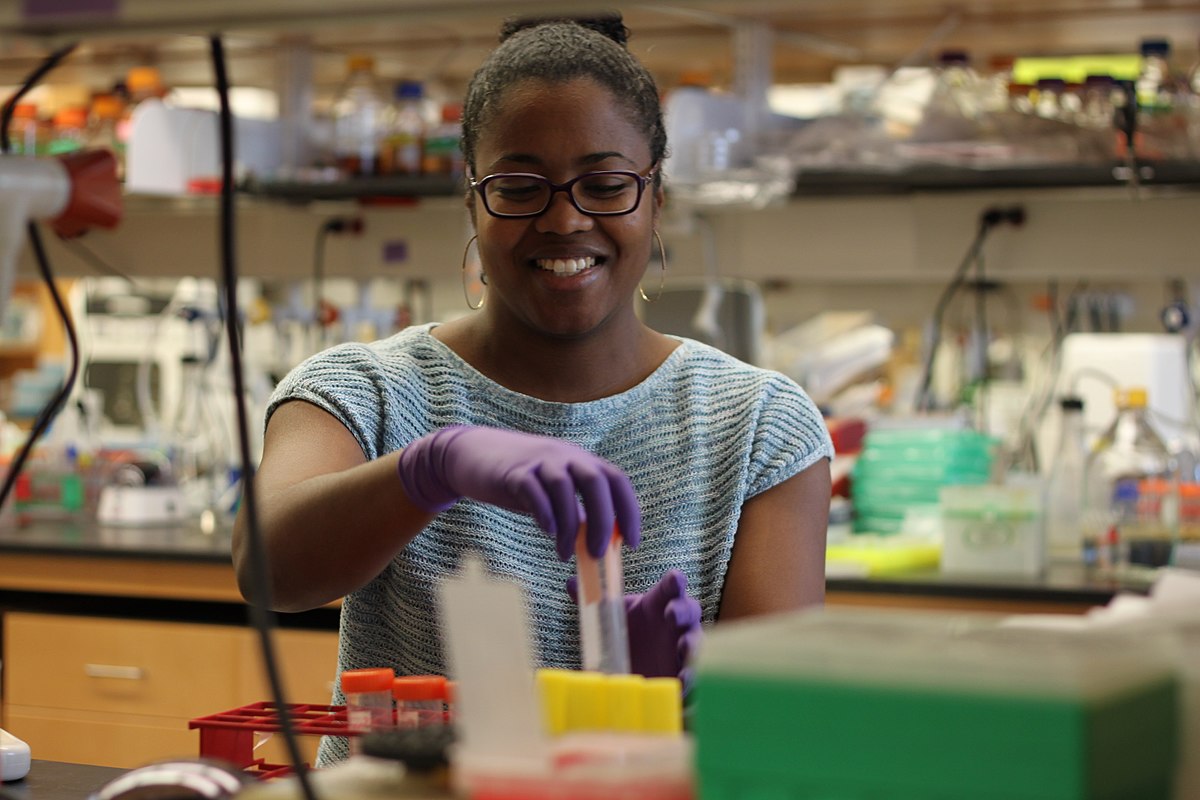
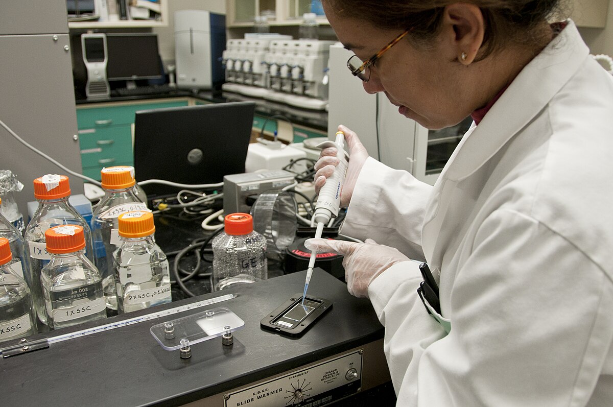
A biochemist wearing glasses and purple gloves smiles while working with test tubes and laboratory equipment. Katherine Stember, CC BY 4.0, via Wikimedia Commons
Beyond the dish
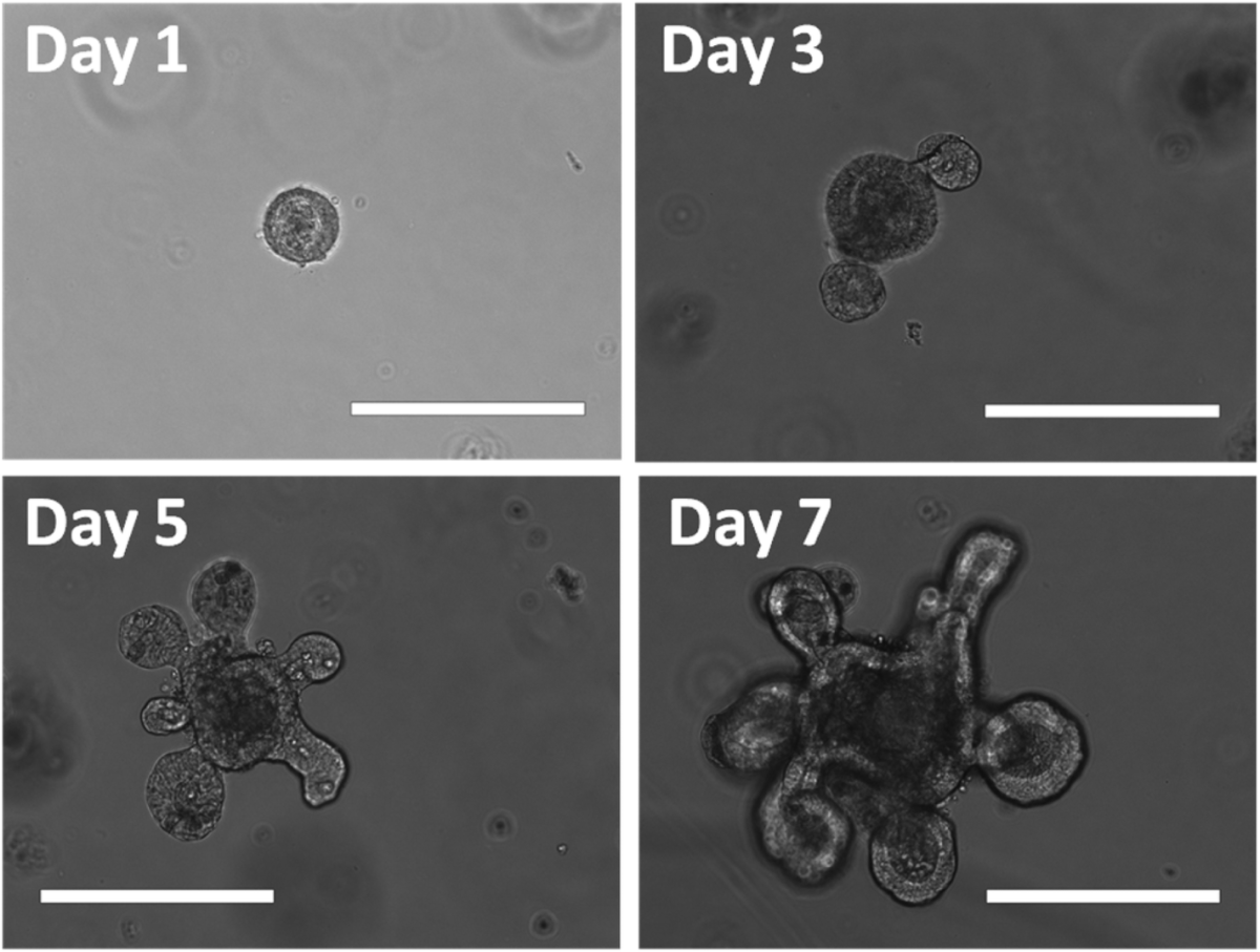
We have moved past simple flat layers of cells. Now, we can grow organoids, miniature, functioning versions of human organs. These tiny tissues allow us to test new chemicals on real human biological systems before a single person is exposed.
Four microscopic images showing the growth of an intestinal organoid (Minigut) over 7 days. Qun Wang, CC BY-SA 4.0, via Wikimedia Commons
It is the ultimate in vitro laboratory.
How cell culture works
Keeping isolated cells alive requires recreating the environment of a living body. Scientists place harvested cells in an incubator set to 37 °C and supply them with a liquid, semi-solid, or solid growth medium such as broth or agar.
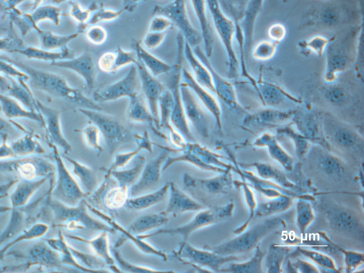
Isolated cells multiply within a nutrient-rich liquid growth medium that mimics the conditions inside a living body. User:Alcibiades, Public domain, via Wikimedia Commons
This medium delivers essential nutrients including amino acids, carbohydrates, vitamins, and minerals, along with hormones, growth factors, and regulated levels of oxygen and carbon dioxide. The environment also maintains a strict pH buffer and osmotic pressure so the cells do not burst or dehydrate.
Depending on the cell type, cells grow either attached to an artificial surface as a single-cell-thick monolayer, called an adherent culture, or floating freely as a suspension culture.
From 2D layers to 3D tissue models
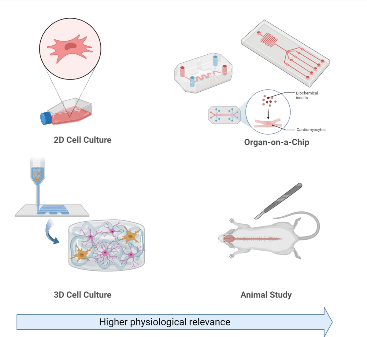
Conventional cell culture grows cells in flat, two-dimensional layers across a dish, causing cells to adopt a flattened shape. However, tissues inside living organisms grow in three dimensions.
Different cultivation methods range from traditional flat 2D layers to 3D scaffolds, organ-on-a-chip setups, and living tissue models. Ayda P, CC BY-SA 4.0, via Wikimedia Commons
Researchers can cultivate cells within gels or polymeric fibrous scaffolds, such as electrospun polycarbonate fibers. When grown on these three-dimensional lattices, cells display a rounded morphology that mirrors how tissues naturally form inside the body.
Vaccine production and medical uses
During the 1940s and 1950s, cell culture advanced to support virology research. Because viruses need living host cells to replicate, growing them in laboratory cell cultures enabled researchers to produce purified viral material for vaccines.
John Franklin Enders, Thomas Huckle Weller, and Frederick Chapman Robbins earned a Nobel Prize for discovering how to grow poliovirus in monkey kidney cell cultures. Jonas Salk used this technique to mass-produce the injectable polio vaccine.
Play the lesson in edgi and the card is yours. It lands on your Map next to the ideas it connects to, and turns from matte to foil to gold as you learn more around it.
What is the difference between primary cells and immortal cell lines?
Primary cells are isolated directly from a donor organism and have a genetically determined lifespan. Immortal cell lines are transformed cells that can divide indefinitely under optimal conditions, such as the HeLa cell line.
Can plant cells be cultured the same way as animal cells?
Plant tissue culture isolates plant cells and places them in a nutrient medium. Because plant cells possess totipotentiality, individual cultured cells have the theoretical capacity to regenerate into an entire plant.