Lactate dehydrogenaseThe enzyme that flips in heart attacks
Lactate dehydrogenase is an enzyme found in almost all living cells that converts pyruvate into lactate and back again. It keeps cells generating energy when oxygen runs low by swapping hydrogen atoms between its fuel molecules and helper cofactors. Because damaged organs leak this enzyme into the bloodstream, doctors use its presence to detect injuries like heart failure.

By the edgi team We find the most surprising true thing about an idea and build a 60-second lesson around it.
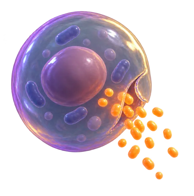
How lactate dehydrogenase manages cellular energy
When oxygen runs short during intense muscle exertion, glycolysis produces pyruvate faster than mitochondria can process it. Lactate dehydrogenase steps in to convert this excess pyruvate into lactate, consuming NADH and regenerating NAD+ in the process.
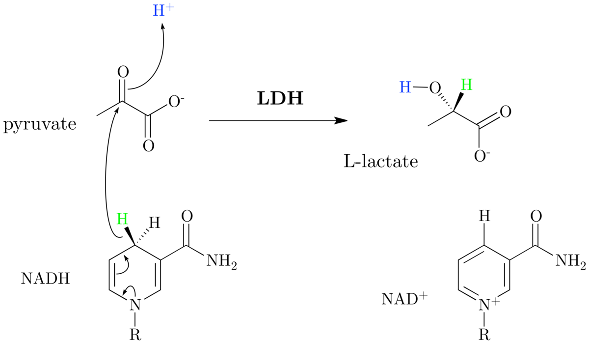
The reaction runs both ways. In the liver, the enzyme performs the reverse step during the Cori cycle, turning lactate back into pyruvate to produce glucose. If lactate builds up to high levels, the enzyme slows itself down through feedback inhibition.
Five tissue-specific isoenzymes
Active lactate dehydrogenase is a tetramer built from combinations of two main building blocks: the M subunit from the LDHA gene and the H subunit from LDHB. These mix to create five distinct forms, labeled LDH-1 through LDH-5.
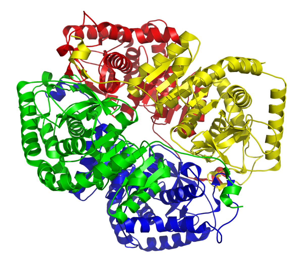
Each form concentrates in specific organs. LDH-1 dominates in the heart and red blood cells, LDH-3 in the lungs, and LDH-5 in the liver and skeletal muscle. Normal blood serum contains mostly LDH-2, but when heart muscle is damaged during a myocardial infarction, it releases large amounts of LDH-1, causing a flipped pattern where LDH-1 levels exceed LDH-2.
Why excess alcohol disrupts blood sugar through LDH
Processing large amounts of ethanol depletes NAD+ and floods liver cells with excess NADH. This imbalance shifts the lactate dehydrogenase reaction equilibrium heavily toward lactate.
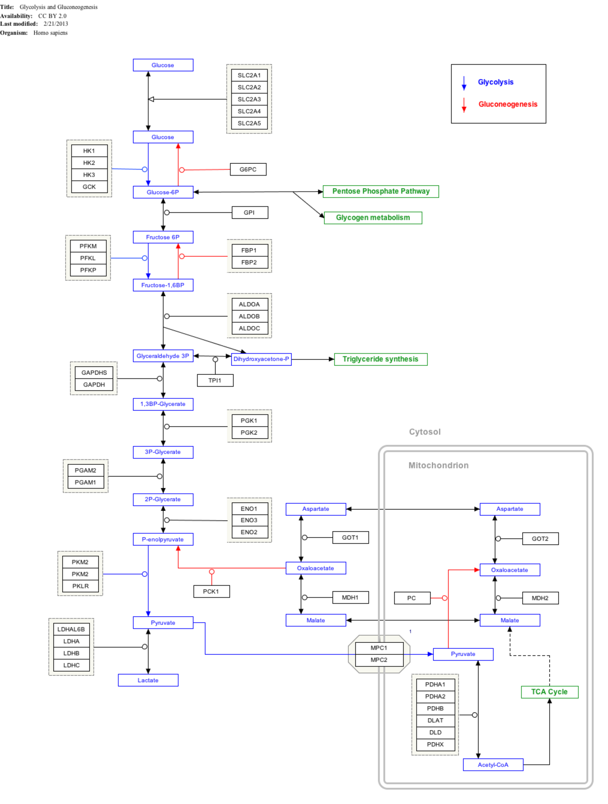
Because the enzyme cannot convert lactate back into pyruvate under these conditions, the liver runs out of the starting material required for gluconeogenesis. In a fasting person who has consumed heavy amounts of alcohol, this reaction stall leads directly to hypoglycemia and lactic acidosis.


Collectible card
Claim the Lactate dehydrogenase card
Play the lesson in edgi and the card is yours. It lands on your Map next to the ideas it connects to, and turns from matte to foil to gold as you learn more around it.
Questions people ask
What is the difference between the H and M subunits of LDH?
The H subunit replaces an alanine residue found in the M subunit with glutamine. This small change allows the H subunit to bind NAD faster and causes its activity to drop fivefold in the presence of acetylpyridine adenine dinucleotide.
Are there other types of LDH subunits beyond H and M?
Yes, mammals also produce LDHC and LDHBx. LDHC is found only in the testes, while LDHBx is a specialized form containing seven extra amino acids that direct it inside peroxisomes.
What amino acid drives the reaction at the active site?
Human lactate dehydrogenase uses a histidine residue at position 193 as its proton acceptor. This residue works alongside arginine and threonine residues that bind the substrate and the coenzyme.


Part of the Set · 9 cards
How Scientists Test What Kills Cells
Before a cancer drug reaches a patient, it has already killed cells in a dish, and someone counted exactly how many.
- Chemotherapy
- Cytotoxicity
- Cell Culture
- Assay
- Necrosis
- Lactate dehydrogenaseReading now
- Dose–response relationship
- IC50
- Therapeutic Window
Where this leads
- ChemotherapyChemotherapy uses systemic drugs to disrupt cell division, stopping rapid cancer growth while balancing toxicity to healthy tissues.
- CytotoxicityLearn what cytotoxicity is, how cells die through apoptosis and necrosis, and how scientists measure cellular toxicity.
- Cell CultureCell culture is the process of growing and maintaining living cells in controlled laboratory environments outside their natural host organism.
- AssayAn assay is a laboratory test that detects, measures, or assesses the activity of a target substance in a sample.