NecrosisHow unprogrammed cell death destroys tissue
Necrosis is the premature death of cells caused by external damage like trauma, toxins, or infection, rather than a planned biological cycle. When a cell suffers necrosis, its outer membrane ruptures and spills toxic enzymes into the surrounding area. This leakage triggers painful inflammation and damages nearby healthy cells, making necrosis harmful and sometimes fatal.
By the edgi team We find the most surprising true thing about an idea and build a 60-second lesson around it.
Most cells die through apoptosis, a tidy, pre-planned suicide where the cell neatly packs itself away. Necrosis is the opposite. It is an accidental, violent death caused by trauma, toxins, or infection.
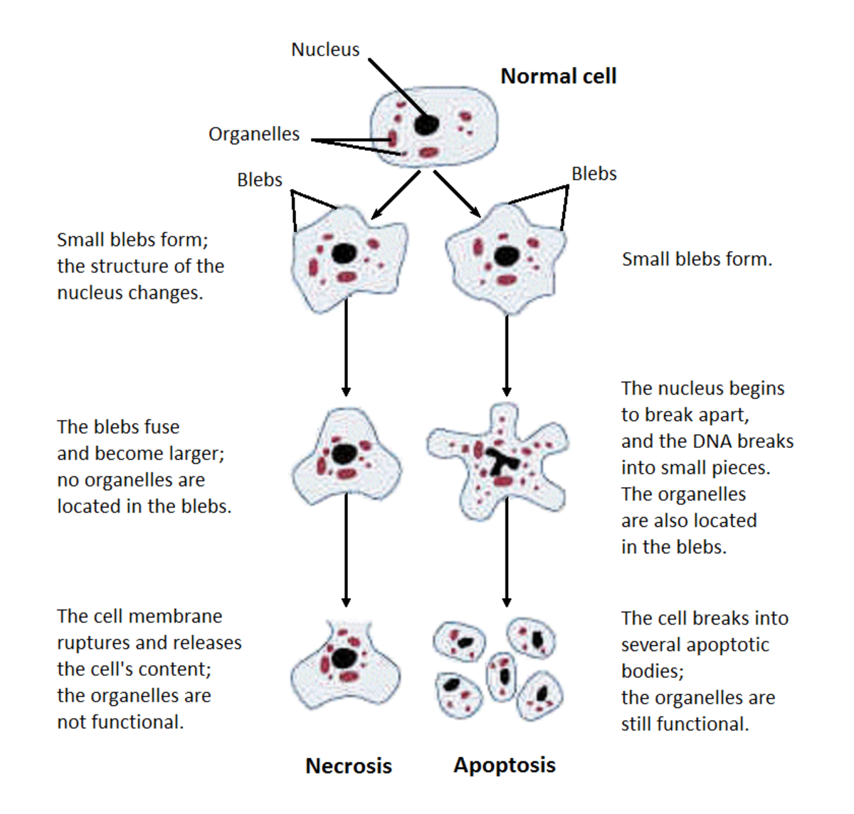
Diagram illustrating the structural changes of cells undergoing necrosis or apoptosis, starting from a normal cell with a nucleus and organelles. National institute on alcohol abuse and alcoholism (NIAAA), Public domain, via Wikimedia Commons
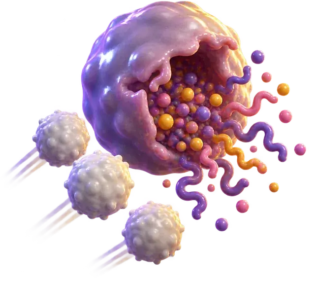
When a cell suffers necrosis, it swells and bursts, spilling its internal contents everywhere.
The biological fire
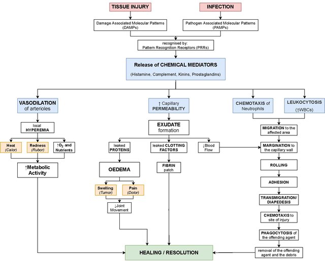
Those spilled contents are toxic. They contain enzymes meant to break down proteins, which now attack neighboring healthy cells. This creates a chain reaction of damage, triggering the heat and swelling we recognize as inflammation.
Flowchart depicting the events of acute inflammation, starting with tissue injury or infection leading to the release of chemical mediators. Gaurav Sinha, CC BY-SA 4.0, via Wikimedia Commons
It is the biological equivalent of a house fire, unlike apoptosis, which is a controlled demolition.
The visible cost
You can see this in action with a bite from a brown recluse spider. The venom cuts off blood flow, causing ischemia. Deprived of oxygen, the tissue begins to die.
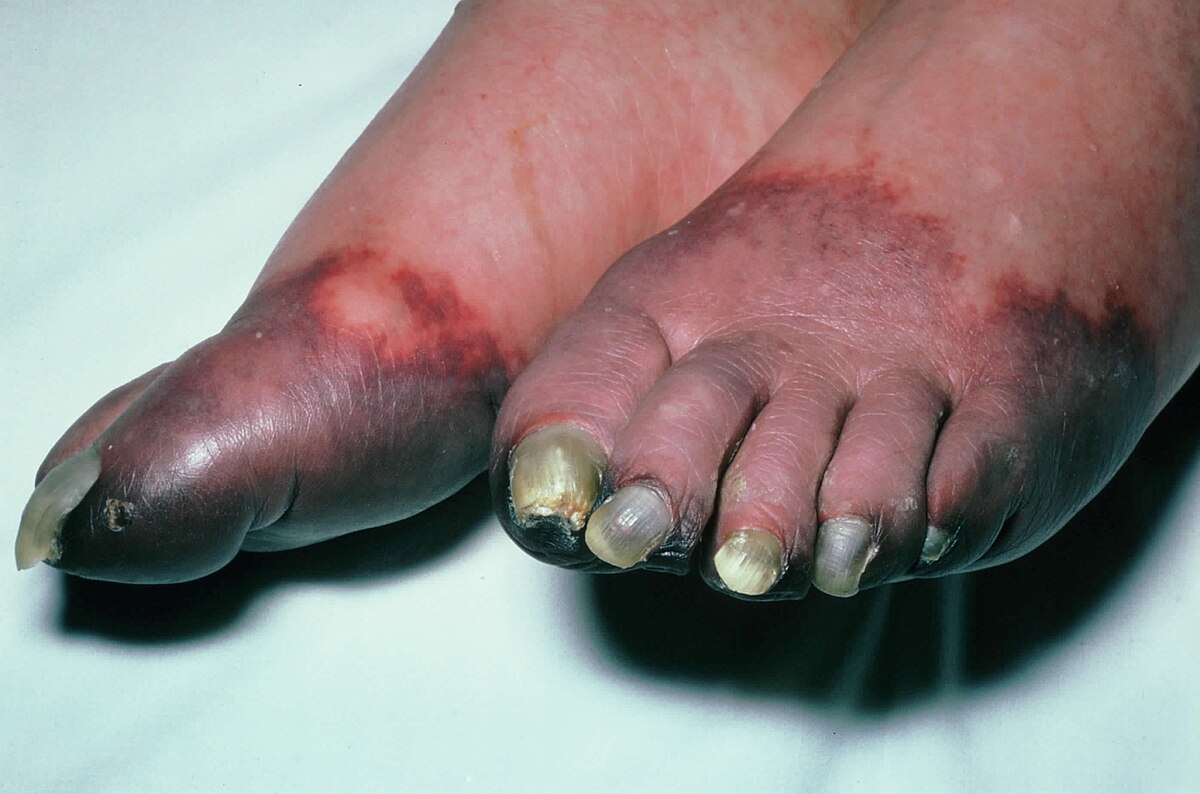
The skin turns dark, liquefies, and rots as the cells literally melt themselves from the inside out. If left unchecked, this spreading rot can lead to gangrene, which sometimes requires surgical removal of tissue to save the body.
Close-up photograph of gangrenous toes on both feet of a diabetic patient. Narraburra, CC0, via Wikimedia Commons
How does necrosis destroy tissue?
Necrosis begins when external forces destroy a cell membrane. Structural signs of this irreversible injury include dense clumping of genetic material and the breakdown of internal organelles. Unlike apoptosis, where a dying cell cleanly packs itself away, necrosis causes the cell to swell and burst.
When the cell bursts, it releases its internal digestive enzymes into the extracellular space. This uncontrolled spill triggers an inflammatory response, calling immune cells like leukocytes and phagocytes to clear the debris. As these immune cells attack the dead tissue, they release damaging substances that cause collateral damage to healthy neighbors, preventing normal healing and causing dead tissue to build up.
The six main patterns of necrosis
Pathologists classify necrosis into six distinct physical patterns based on how the dead tissue looks and behaves. Coagulative necrosis happens in low-oxygen environments like the heart or kidneys, leaving a firm, gel-like structure of denatured proteins. Liquefactive necrosis occurs when cells turn into a viscous liquid mass, known as pus, which is typical of bacterial infections and brain infarcts.
Other patterns include caseous necrosis, which forms a crumbly, cheese-like debris seen in tuberculosis infections, and fat necrosis, where leaked pancreatic enzymes break down fats into chalky white calcium deposits. Gangrenous necrosis affects limbs starved of oxygen, and fibrinoid necrosis occurs when immune complexes and fibrin collect inside damaged blood vessel walls.
Medical consequences and treatment
Left untreated, necrotic tissue accumulates into conditions like gangrene, where limbs mummify or rot from severe lack of oxygen. Spider bites from the brown recluse in the United States and the Chilean recluse in South America cause localized ischemia that can quickly develop into spreading necrosis.
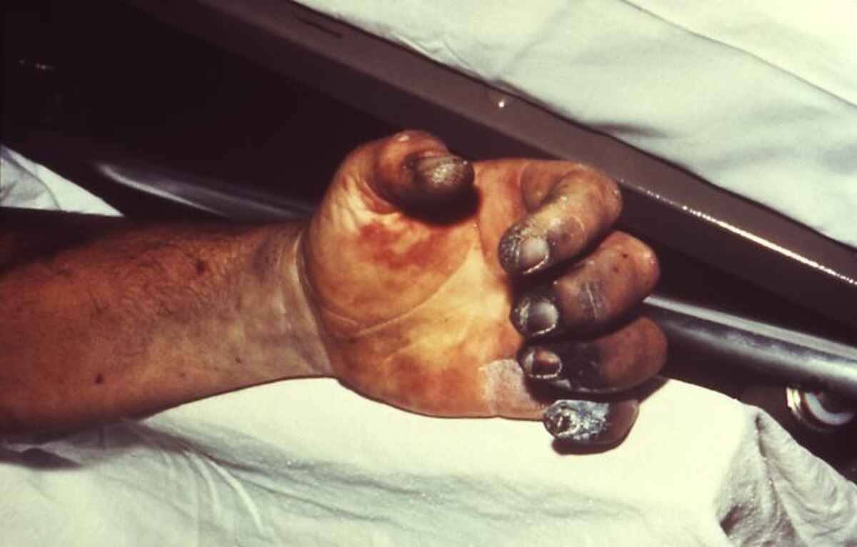
Blackened tissue shows how severe infection and loss of blood supply cause dead flesh to accumulate on a limb. CDC/Dr. Jack Poland, Public domain, via Wikimedia Commons
Because necrotic tissue harbors decomposing debris and harmful enzymes that actively poison adjacent healthy flesh, the body cannot easily heal on its own. Doctors frequently must remove the dead tissue through a surgical procedure called debridement to stop the damage from spreading.
Play the lesson in edgi and the card is yours. It lands on your Map next to the ideas it connects to, and turns from matte to foil to gold as you learn more around it.
The term necrosis comes from the Ancient Greek word for death. It entered medical usage in the mid-19th century and is widely attributed to German pathologist Rudolf Virchow.
What is the difference between dry gangrene and wet gangrene?
Dry gangrene is a type of coagulative necrosis where tissue resembles mummified skin due to lack of blood flow. Wet gangrene occurs when that dead tissue suffers a bacterial infection, turning it into liquefactive necrosis.
Do all spider bites cause necrosis?
No, most do not. In the United States, only the venom of the brown recluse reliably causes necrosis, while claims about yellow sac and hobo spiders causing it have not been substantiated.