P53The cell inspector that triggers self-destruction
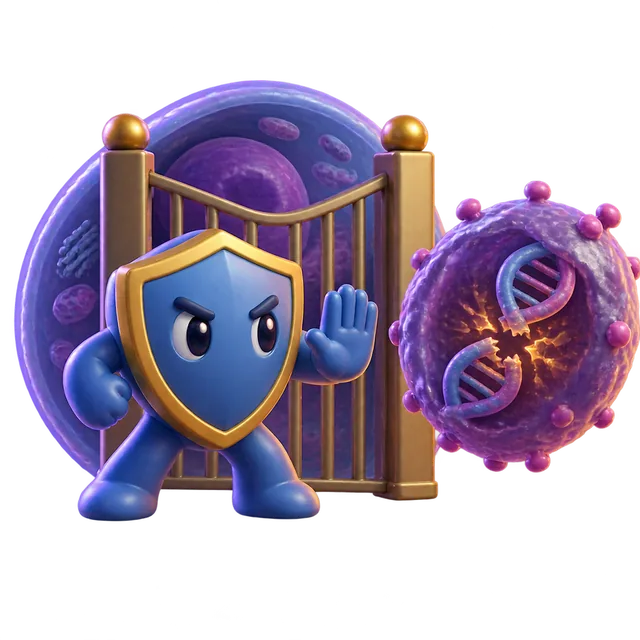
P53 is a protein that acts as a quality control inspector inside your cells, binding to DNA to control which genes get turned on. It prevents cancer by pausing the cell cycle to allow DNA repair or by ordering hopelessly damaged cells to self-destruct. More than half of all human cancers involve a mutated or inactive version of this single protein.
By the edgi team We find the most surprising true thing about an idea and build a 60-second lesson around it.
Every day, your cells make tiny copying errors that could turn into tumors. A single protein called p53 acts as a quality-control inspector, freezing the cell's life cycle to fix the damage.
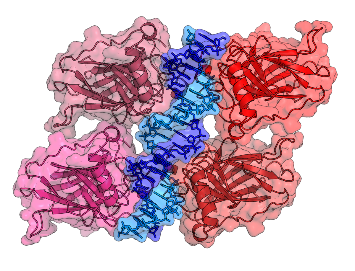
Crystal structure of four p53 DNA-binding domains, forming a homo-tetramer, bound to a DNA double helix. Richard Wheeler (Zephyris), CC BY-SA 3.0, via Wikimedia Commons
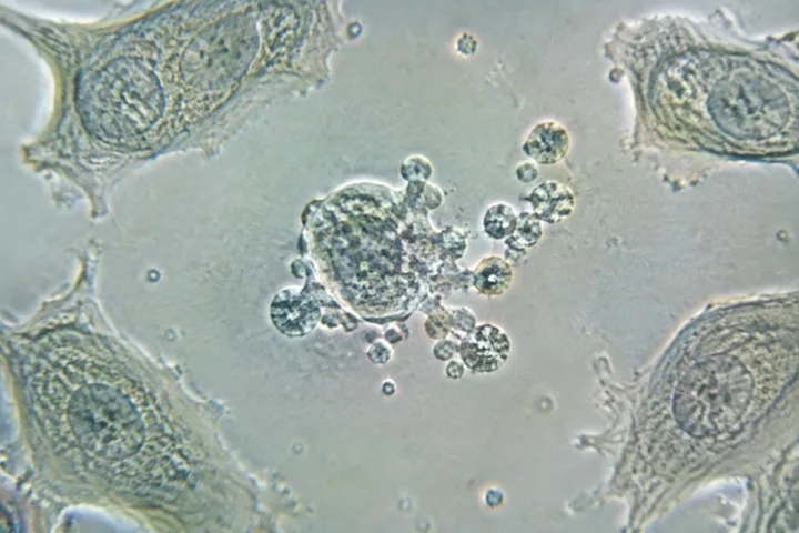
If the error is too severe, p53 triggers apoptosis, ordering the damaged cell to self-destruct.
Mistaken identity
When researchers first discovered p53 in 1979, they misidentified it as a cancer-causing agent. It took a decade of confusing data to realize they had been studying a mutated, broken version of the protein.
In 1989, scientist Bert Vogelstein proved the healthy version was actually a vital tumor suppressor.
The elephant exception
Because elephants are massive and live long lives, they should theoretically contract cancer at staggering rates. Instead, evolutionary biologist investigations revealed they carry twenty copies of the TP53 gene.
When their cells detect damaged DNA, their abundance of p53 ruthlessly snuffs out the threat before it spreads.
How p53 protects cells from DNA damage
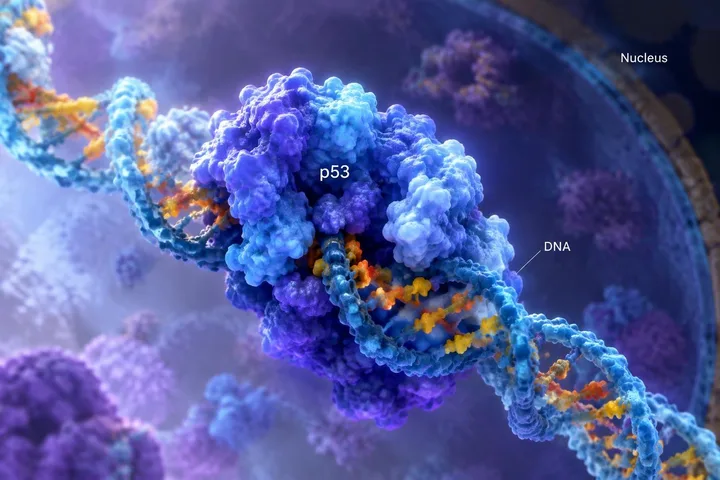
A healthy p53 protein operates as a transcription factor, meaning it latches onto specific sections of DNA to switch protective genes on or off. When a cell experiences genetic damage, p53 halts cell division at the G1/S checkpoint. This pause gives internal repair proteins the time they need to fix errors before the cell duplicates.
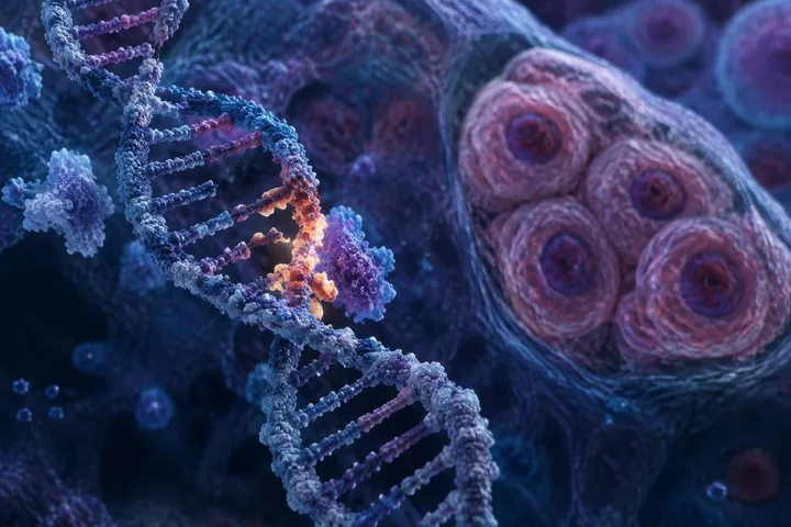
The p53 protein binds directly to the DNA double helix to turn on repair genes or trigger cell death. Thomas Splettstoesser, CC BY-SA 3.0, via Wikimedia Commons
If the genetic damage is too severe to mend, p53 switches strategies and activates pro-apoptotic genes to trigger programmed cell death. It also steps in during cellular senescence, the permanent retirement of aging cells caused by shortened telomeres. By eliminating unstable cells, p53 stops rogue mutations from multiplying into tumors.
The physical structure of the protein
P53 does not work as a lone molecule. Instead, it functions as a tetramer, a four-part unit made of two interlocking pairs called dimers. Dimers assemble while being built on ribosomes, then join together post-translationally into the active four-part complex.
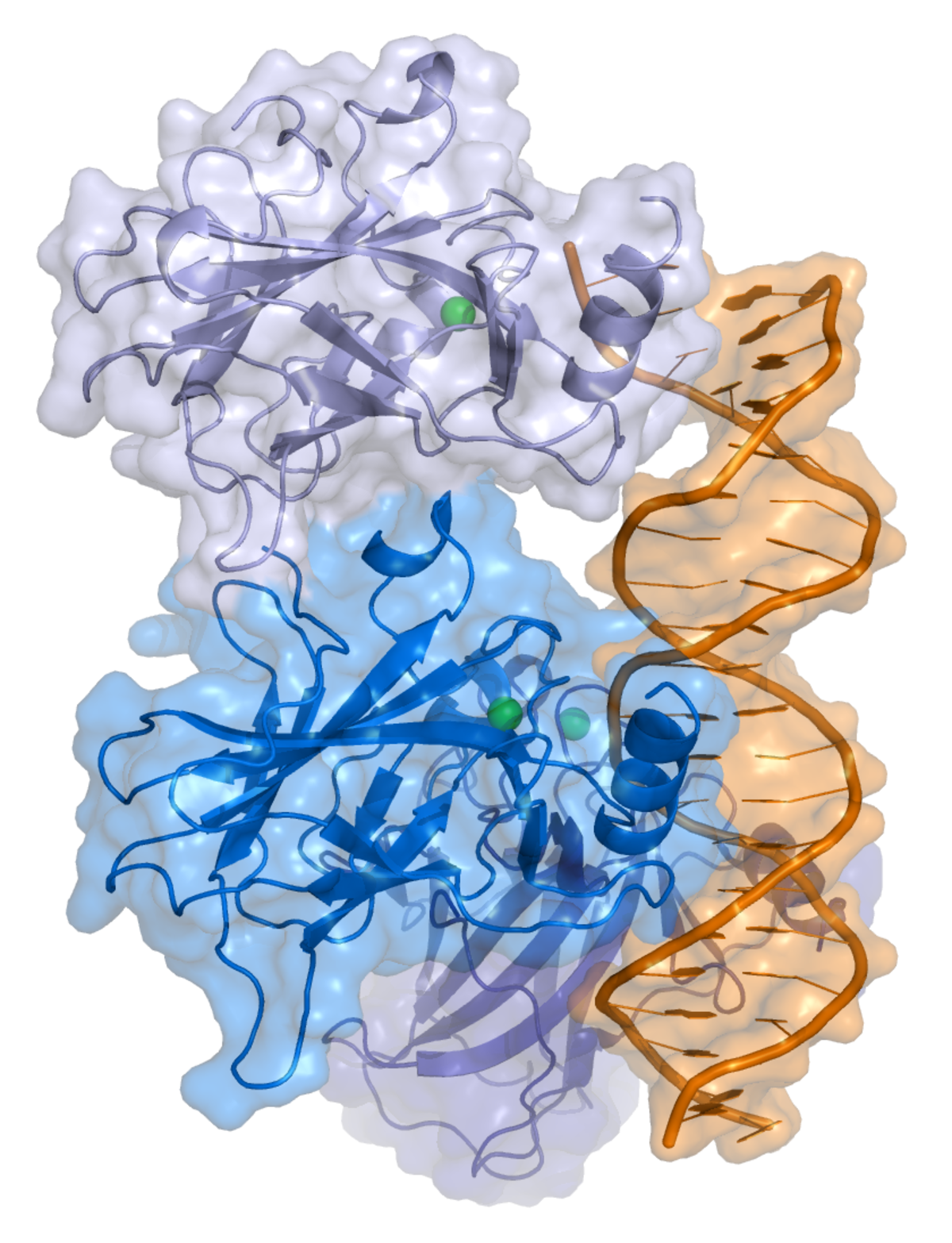
The full-length p53 protein contains seven functional domains, including an acidic transactivation domain that turns on target genes, a central DNA-binding domain holding a zinc atom, and an oligomerization domain that locks the tetramer together. Most cancer-causing mutations strike directly at the DNA-binding domain, ruining the protein's ability to grab onto target genes.
Where the TP53 gene lives in the human genome
In humans, the TP53 gene sits on the short arm of chromosome 17 at band 17p13.1. The gene spans 20 kilobases of genetic sequence and contains a long non-coding intron of 10 kilobases. Beyond the standard full-length protein, human cells use alternative splicing, internal promoters, and multiple start sites to manufacture at least 12 distinct variations, or isoforms.
Human chromosome 17 contains the TP53 gene on its upper arm at band 17p13.1. Own work, Public domain, via Wikimedia Commons
Modern humans carry over 1,000 mutated variations in the gene that are absent in Neanderthal populations. Genomic studies show that the vast majority of these protein-altering variants emerged between 5,000 and 10,000 years ago.
Test yourself
When researchers first found p53, did they think it caused or stopped cancer?
They thought it caused cancer. Researchers initially studied a mutated, broken version of p53, tricking them into thinking the normal protein promoted tumor growth for a decade.
The p53 protein exists to accelerate cell growth for faster tissue repair.
False. P53 is not a growth accelerator; it is a quality-control inspector that pauses the cell cycle to fix DNA damage, preventing mutations from becoming cancerous.
Play the lesson in edgi and the card is yours. It lands on your Map next to the ideas it connects to, and turns from matte to foil to gold as you learn more around it.
Why is the protein called p53 if its weight is 43.7 kDa?
Early laboratory measurements using SDS-PAGE gel electrophoresis made the protein appear to weigh 53 kilodaltons. Its high content of the amino acid proline slows its movement through testing gels, creating the illusion of extra mass.
What are p53 isoforms?
Isoforms are 12 different structural versions of p53 produced from the same gene, ranging in size from 3.5 to 43.7 kilodaltons. Some isoforms lack the specific regions needed to trigger cell death, giving different tissues varied ways to regulate cell survival.