ADCC: how immune cells destroy antibody-flagged targets
Antibody-dependent cellular cytotoxicity is an immune defense where specialized immune cells destroy target cells that have been flagged by antibodies. Instead of relying solely on floating immune proteins to punch holes in a target, it requires a live immune cell to recognize the antibody handle and release lethal chemicals. This pathway helps the body clear virus-infected cells, eliminate tumors, and destroy large parasites.
By the edgi team We find the most surprising true thing about an idea and build a 60-second lesson around it.
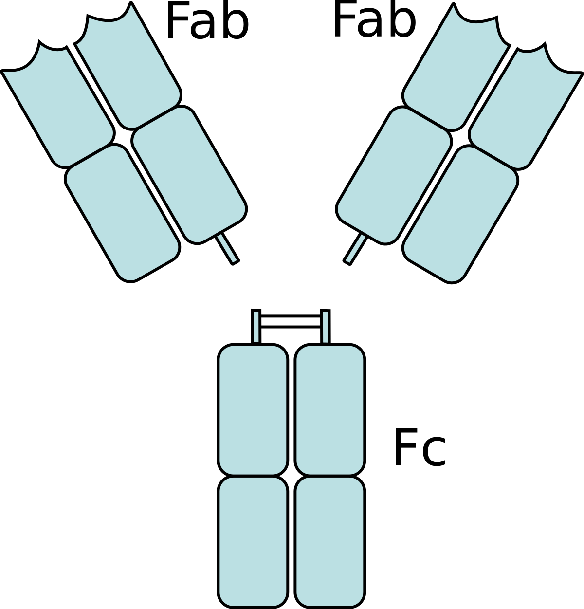
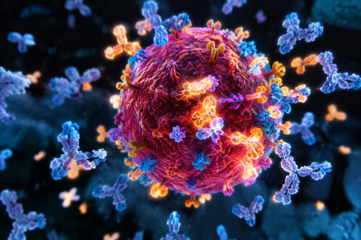
Antibodies are shaped like tiny Y letters. The forked tips grab onto specific surface antigens on a virus or tumor cell. The base of the Y is called the fragment crystallizable region, or Fc region for short.
Diagram showing the two Fab (fragment, antigen-binding) regions and one Fc (fragment, crystallizable) region of an antibody. 2fab fc.png: User Je at uwo on en.wikipedia / derivative work: Vezixig, Public domain, via Wikimedia Commons
That tail acts as a molecular handle, waiting for an immune cell to grab it.
The hit squad
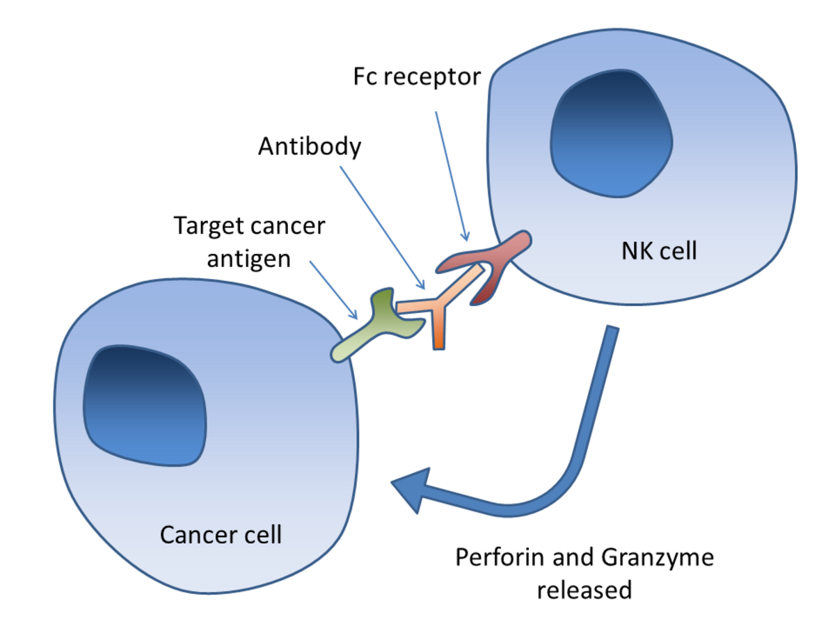
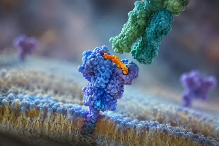
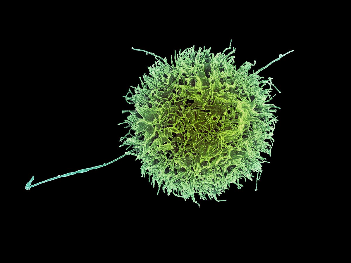
Natural killer cells patrol the body looking for cells that have been marked for destruction. They carry specialized proteins on their surface called Fc receptors that lock directly onto the antibody tails.
Diagram of antibody-dependent cell-mediated cytotoxicity (ADCC) illustrating the interaction between a natural killer (NK) cell and a cancer cell. Simon Caulton, CC BY-SA 3.0, via Wikimedia Commons
Once an Fc receptor engages a bound antibody, the natural killer cell is given the green light to attack.
The killing blow
Unlike other immune defenses that punch holes in targets using proteins alone, this mechanism requires a living effector cell. The natural killer cell releases toxic enzymes like perforin and granzymes directly onto the bound target.
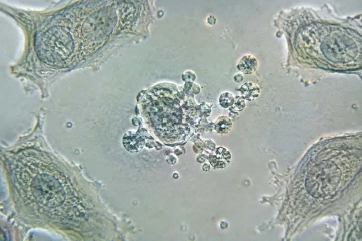
Those chemicals force the doomed cell to self-destruct from the inside out.
How effector cells trigger cell death
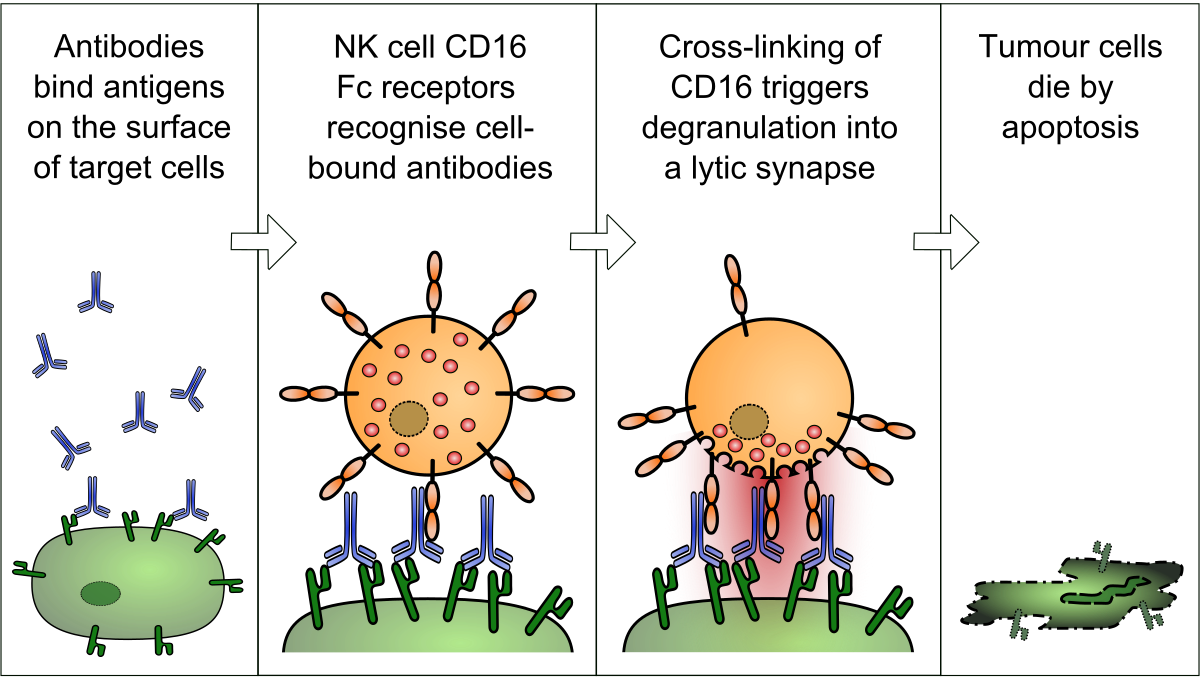
The classic pathway relies on natural killer cells interacting with immunoglobulin G antibodies. When a cell is infected by a virus, viral proteins appear on its outer membrane. Specific IgG antibodies bind to these surface antigens, leaving their tail regions pointing outward.
An effector cell binds to antibody tails on a target cell to trigger membrane lysis. Satchmo2000, CC BY-SA 3.0, via Wikimedia Commons
Natural killer cells carry surface receptors called Fc receptors, most commonly CD16 or FcγRIII. These receptors lock onto the exposed antibody tails. Once engaged, the natural killer cell releases toxic enzymes, including perforin and proteases called granzymes. These chemicals cause lysis, breaking down the target cell membrane to stop the virus from spreading.
Destroying large parasites
Certain invaders are too massive for immune cells to engulf through phagocytosis. Parasitic worms known as helminths have a tough outer structure that resists chemicals released by macrophages and neutrophils.
Eosinophils overcome this barrier using a specific form of cytotoxicity. IgE antibodies coat the surface of the parasite, and specialized Fc receptors on the eosinophil bind to these antibodies. This direct contact signals the eosinophil to degranulate, releasing its contents to damage the large parasite.
Medical uses in cancer therapy
This cytotoxic mechanism plays a central role in several monoclonal antibody treatments for cancer. In mouse experiments, therapies against solid tumors using antibodies like trastuzumab and rituximab rely on this response for their therapeutic action. It is also an active mechanism in daratumumab treatments for multiple myeloma.
In laboratory research, scientists measure the strength of this response using release assays. Target cells are loaded with radioactive isotopes such as chromium-51 or fluorescent dyes like calcein. When effector cells lyse the antibody-coated targets, the released markers are measured to quantify how effectively the cells were destroyed.
Test yourself
In antibody-dependent cellular cytotoxicity, the antibody itself acts as the lethal agent that punches holes in the target cell's membrane.
False. Antibodies only serve as molecular tags. The actual killing is performed by natural killer cells, which release toxic enzymes after binding to the antibody's tail.
Claim the Antibody-dependent cellular cytotoxicity card
Play the lesson in edgi and the card is yours. It lands on your Map next to the ideas it connects to, and turns from matte to foil to gold as you learn more around it.
The complement system can lyse target cells using circulating blood proteins alone without requiring an immune cell. In contrast, antibody-dependent cellular cytotoxicity strictly requires a living effector cell, such as a natural killer cell or eosinophil, to deliver the killing blow.
Why is ADCC more common against viruses than bacteria?
IgG antibodies bind readily to viral antigens displayed on host cell surfaces, allowing effector cells to destroy the infected cell. Against bacterial infections, antibodies mainly neutralize bacterial toxins or coat infected host cells rather than triggering direct cellular destruction of the prokaryotes.
Which immune cells can act as effector cells?
Natural killer cells are the classic effector cells, but macrophages, neutrophils, and eosinophils can also perform this function. For example, eosinophils use it to attack large helminth parasites coated with IgE antibodies.