The hit squad
Your immune system includes specialized cells that hunt down and destroy your own cells when they are hijacked by viruses or turn cancerous. They are called killer T cells, and they carry a literal license to execute damaged tissue.
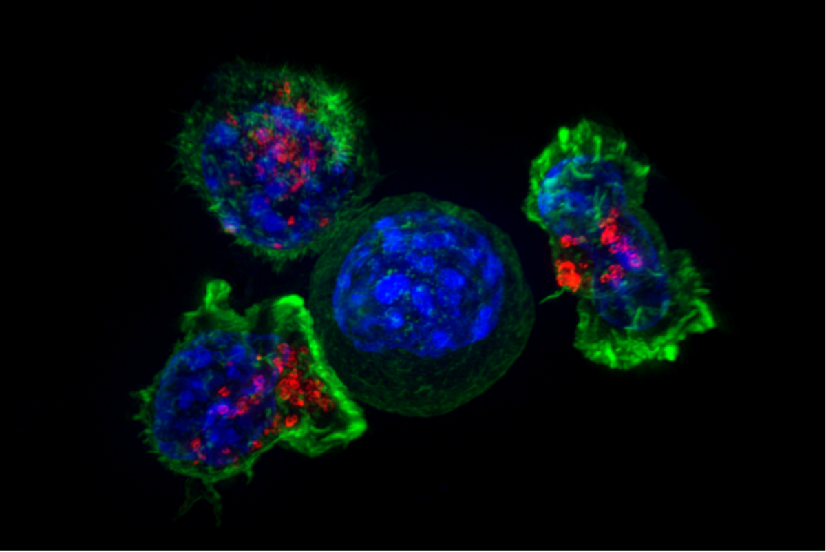
Killer T cells are white blood cells that find and destroy your own body cells when they turn cancerous or get infected by viruses and bacteria. They patrol the bloodstream and read chemical fragments displayed on cell surfaces to spot threats. Once a target is confirmed, they latch on and trigger programmed cell suicide to wipe out the damaged cell.

By the edgi team We find the most surprising true thing about an idea and build a 60-second lesson around it.
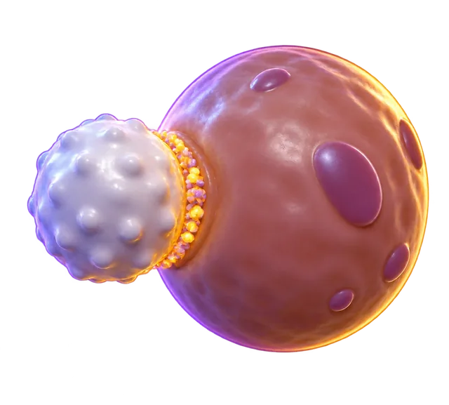
Your immune system includes specialized cells that hunt down and destroy your own cells when they are hijacked by viruses or turn cancerous. They are called killer T cells, and they carry a literal license to execute damaged tissue.
Killer T cells patrol the bloodstream looking for a specific antigen displayed on the surface of target cells. They check these chemical fingerprints against their own T-cell receptors before launching an attack.
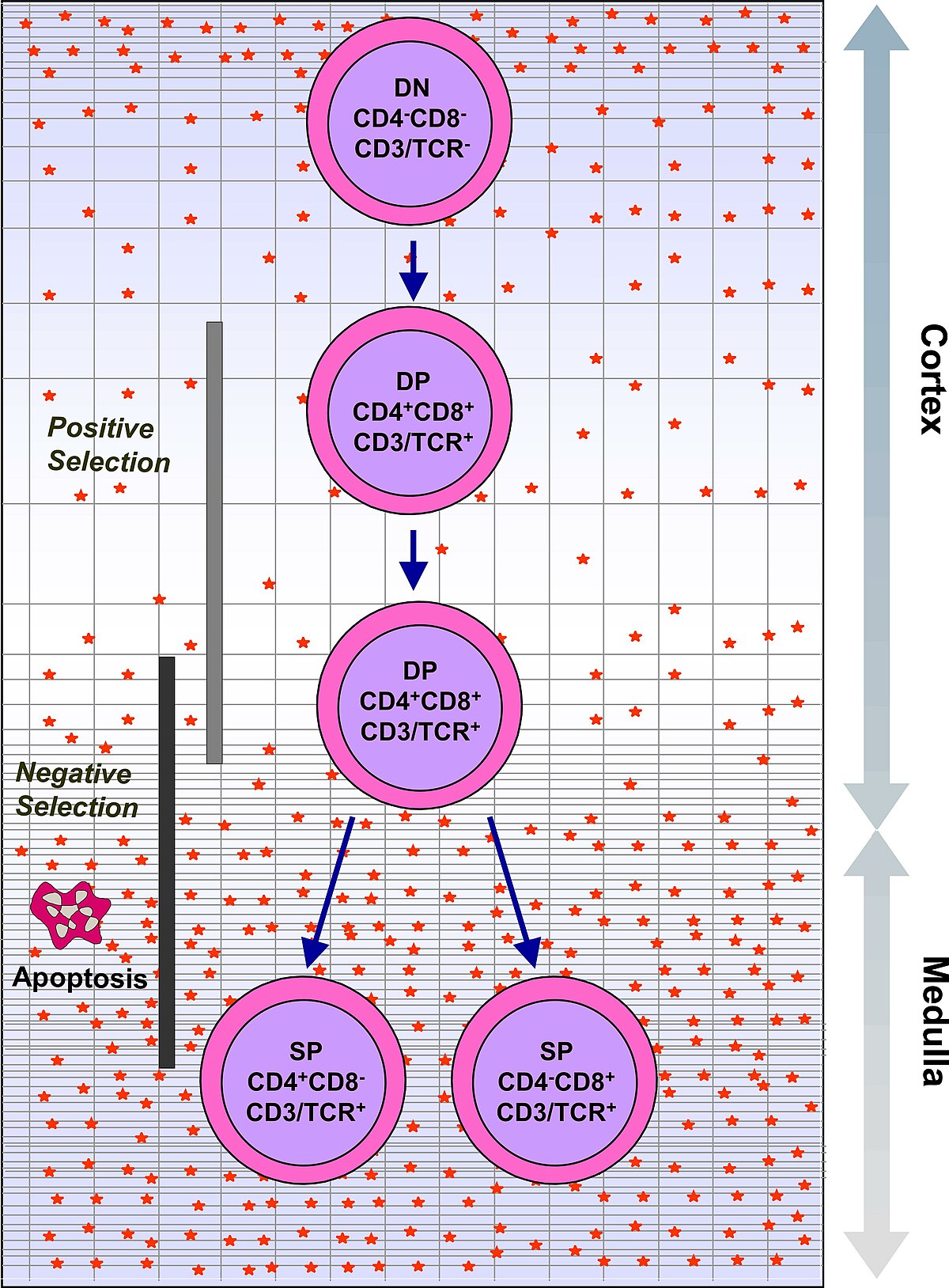
To prevent them from attacking healthy tissue, immature T cells endure a brutal screening process in the thymus where 98% are killed. The survivors learn to recognize MHC class I molecules, the protein stands healthy cells use to show what is happening inside them.
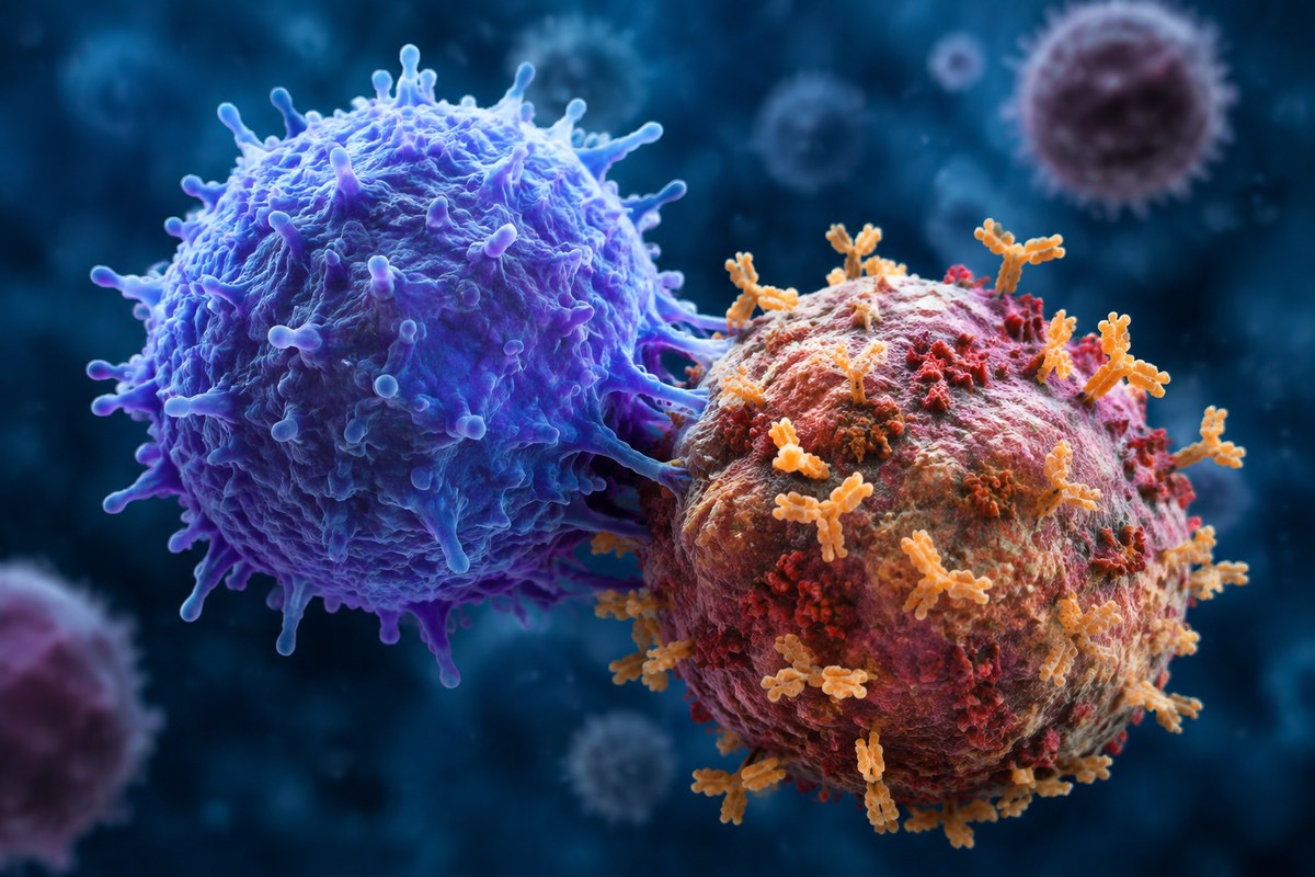
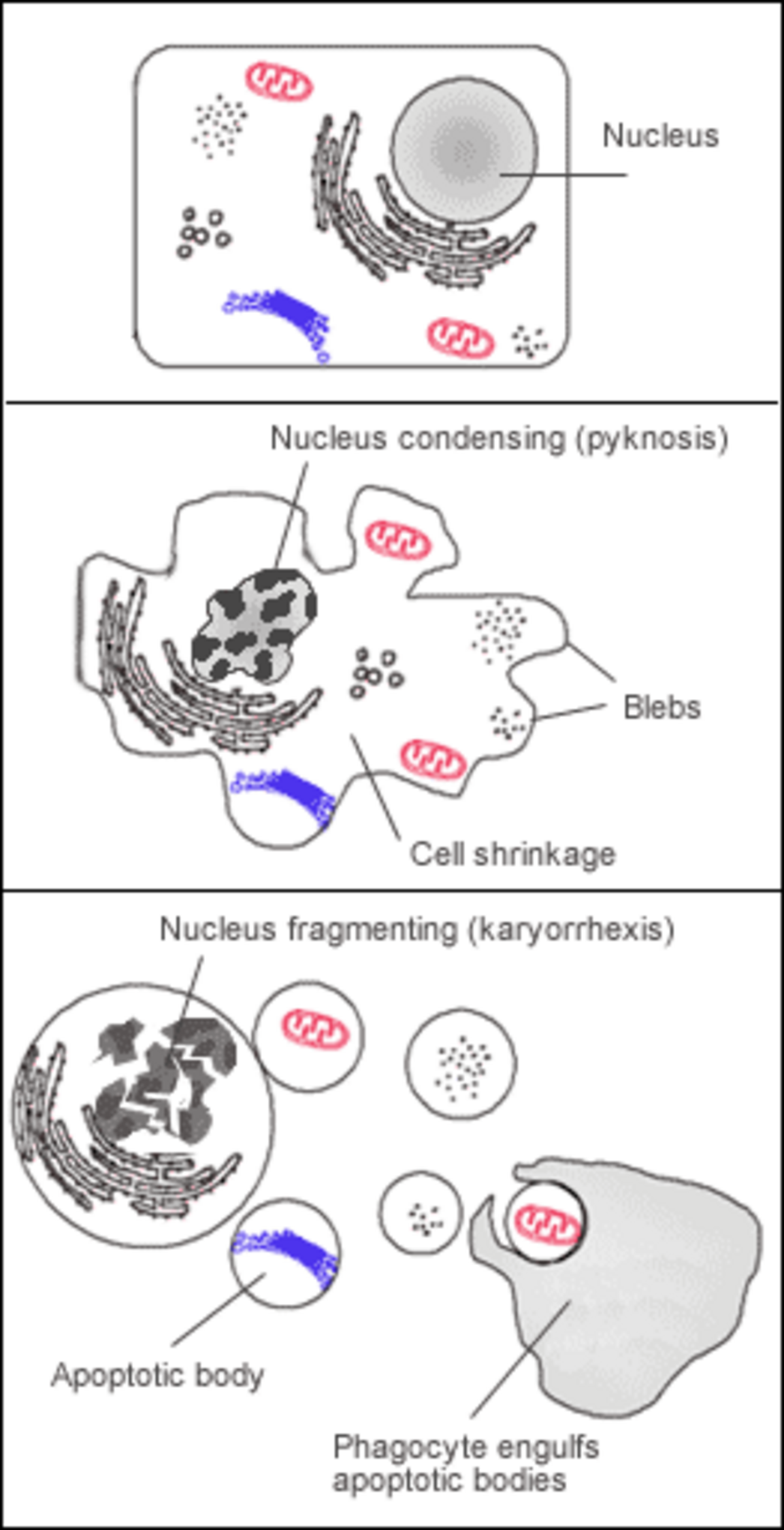
When a match is found, the T cell forms a tight seal called an immunological synapse with the rogue cell. It releases toxic proteins that trigger apoptosis, forcing the infected cell to commit programmed suicide.
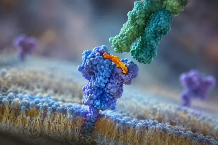
A healthy human cell continuously displays bits of internal proteins on its surface using molecular stands called class I MHC molecules. Killer T cells, formally known as CD8+ T cells, carry specialized T-cell receptors (TCRs) that check these displayed protein pieces, called antigens. A glycoprotein on the killer T cell called CD8 binds to the constant portion of the class I MHC molecule, locking the two cells tightly together while the receptor checks the antigen.
If the surface antigen matches what the T-cell receptor is built to recognize, the killer T cell activates and forms a tight immunological synapse with the rogue cell. It releases toxic proteins that force the infected or cancerous target to destroy itself through apoptosis.
The human body has fewer than 30,000 genes, yet it must identify over a billion potential antigens. To solve this, developing white blood cells shuffle their TCR gene segments in the bone marrow through V(D)J recombination, creating millions of random variations.
Immature cells travel to the thymus, an organ that requires iodine to develop and function. There, double-positive cells displaying both CD4 and CD8 co-receptors face two strict screening tests. Positive selection preserves cells that can properly bind to self MHC molecules. Negative selection immediately kills off cells that bind too strongly to the body's own healthy antigens. The surviving cells drop CD4, keep CD8, and emerge as mature killer T cells ready to respond to class I MHC targets.
Killer T cells protect the body mainly by killing which cells?
The body's infected or cancerous cells. Killer T cells destroy the body's own infected or cancerous cells, limiting threats that have taken over or altered those cells.
How do Killer T cells determine which body cells to destroy?
They scan MHC class I molecules for antigens.. Killer T cells do not look for generic damage. They specifically check MHC class I molecules to identify if the internal proteins are infected or cancerous.
Apoptosis, or programmed cell death, is the primary mechanism by which Killer T cells eliminate their targets.
True. By triggering a controlled suicide mission in the target cell, the T cell ensures the pathogen doesn't spill out and spread.
Collectible card
Play the lesson in edgi and the card is yours. It lands on your Map next to the ideas it connects to, and turns from matte to foil to gold as you learn more around it.
A naive T cell is a mature cell that has never encountered its matching antigen. Once activated by an antigen match, it turns into an effector T cell capable of killing targets. After clearing the threat, some survive in a resting state as memory T cells, ready to reactivate quickly if the same antigen reappears.
Yes. Activated killer T cells can release cytokines such as TNF-alpha and IFN-gamma. These signaling molecules provide additional antitumour and antimicrobial effects alongside direct cell killing.
While most killer T cells carry alpha and beta receptor chains, some cells in epithelial tissues like the gut carry gamma and delta chains instead. These gamma-delta T cells can recognize non-protein antigens, stress proteins, and microbial toxic shock proteins without needing antigens presented on MHC molecules.
Part of the Set · 8 cards
Almost every cell in your body carries an ID badge. Show the wrong one, or none, and your own immune system kills it.
Learn the whole SetLesson complete
You finished the lesson. In edgi, the card goes on your Map next to everything else you learn.