Major histocompatibility complexThe cellular ID system
The Major Histocompatibility Complex (MHC) is a cluster of genes that produce surface proteins found on almost all cells in jawed vertebrates. These proteins act like display windows, holding up fragments of internal proteins so passing immune cells can check what is happening inside. By showing whether a cell is healthy or infected, MHC helps determine organ transplant compatibility and defends the body against viruses and cancers.
By the edgi team We find the most surprising true thing about an idea and build a 60-second lesson around it.
Every cell in your body constantly displays tiny fragments of its internal proteins on its surface. This genetic system is called the Major Histocompatibility Complex, or MHC.
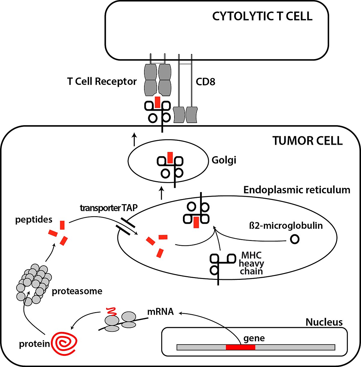
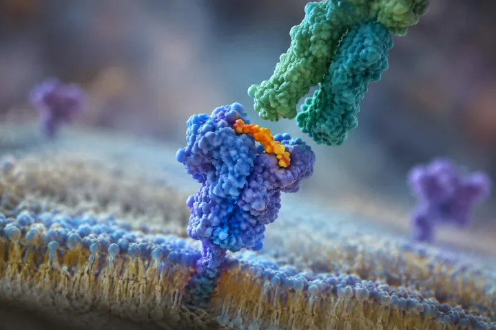
Diagram showing the processing of tumor antigens recognized by CD8+ T cells within a tumor cell. UCLLICRBRU, CC BY-SA 4.0, via Wikimedia Commons
It acts as a continuous identification checkpoint, allowing roaming immune cells to inspect what is happening inside.
Friend or foe
If a cell is hijacked by a virus or turns cancerous, the proteins it displays will change. T cells inspect these surface displays using specialized receptors.
If the ID looks wrong or missing, the immune system assumes the cell is dangerous and destroys it.
The transplant puzzle
In the 1940s, geneticist George Snell discovered these exact genes while studying why mouse tumor transplants mysteriously failed. He realized the immune system rejects foreign tissue not because it is weak, but because the donor's MHC tags do not match.
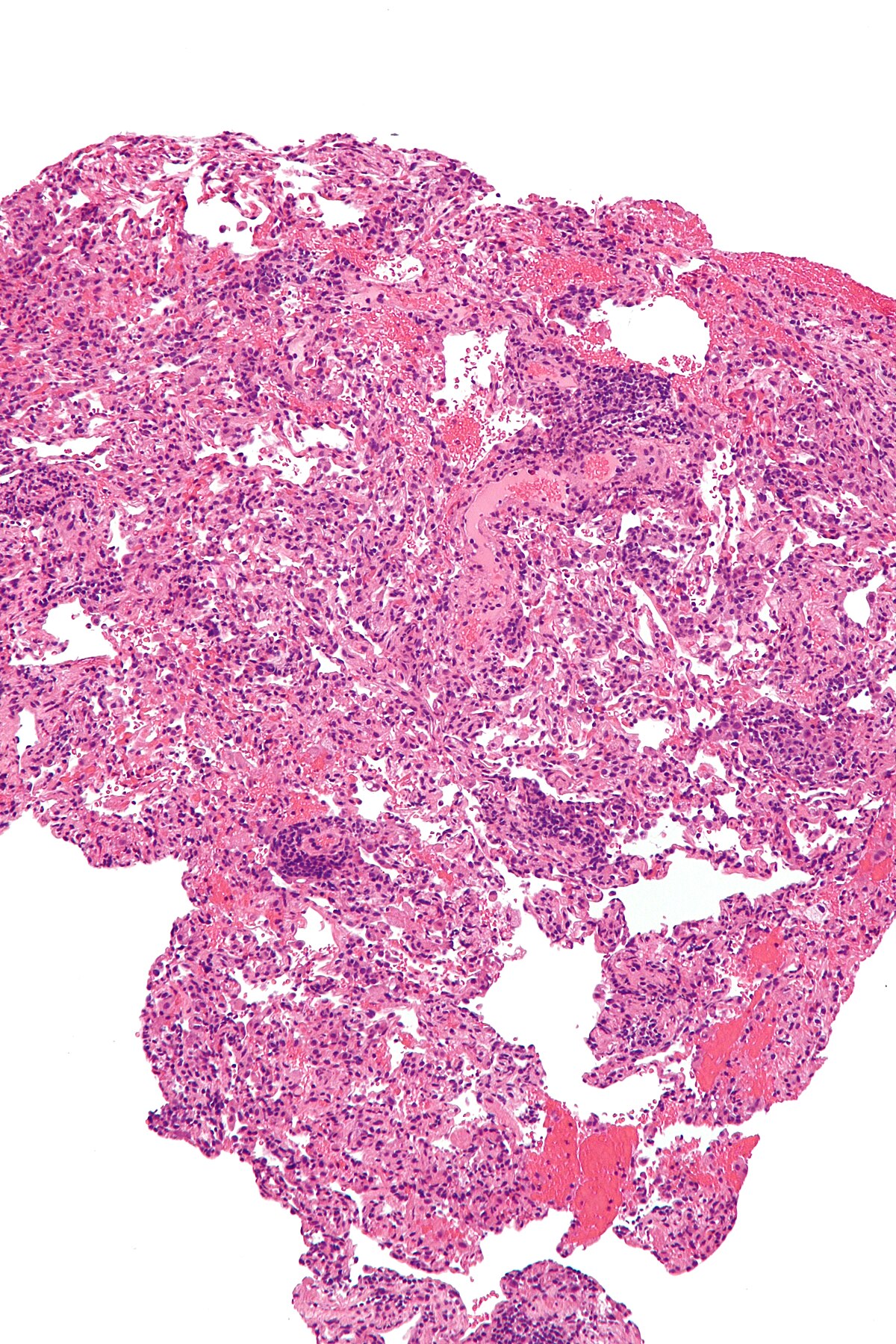
A micrograph showing lung transplant rejection, with a lung biopsy stained with H&E. Nephron, CC BY-SA 3.0, via Wikimedia Commons
This strict vetting is also why Autoimmune Disorders happen when the security system mistakes healthy tissue for an invader.
How MHC molecules present antigens
Inside every cell, protein molecules are constantly made and broken down. MHC molecules take small protein fragments, called epitopes, and carry them to the outer cell surface for inspection by white blood cells called T cells.
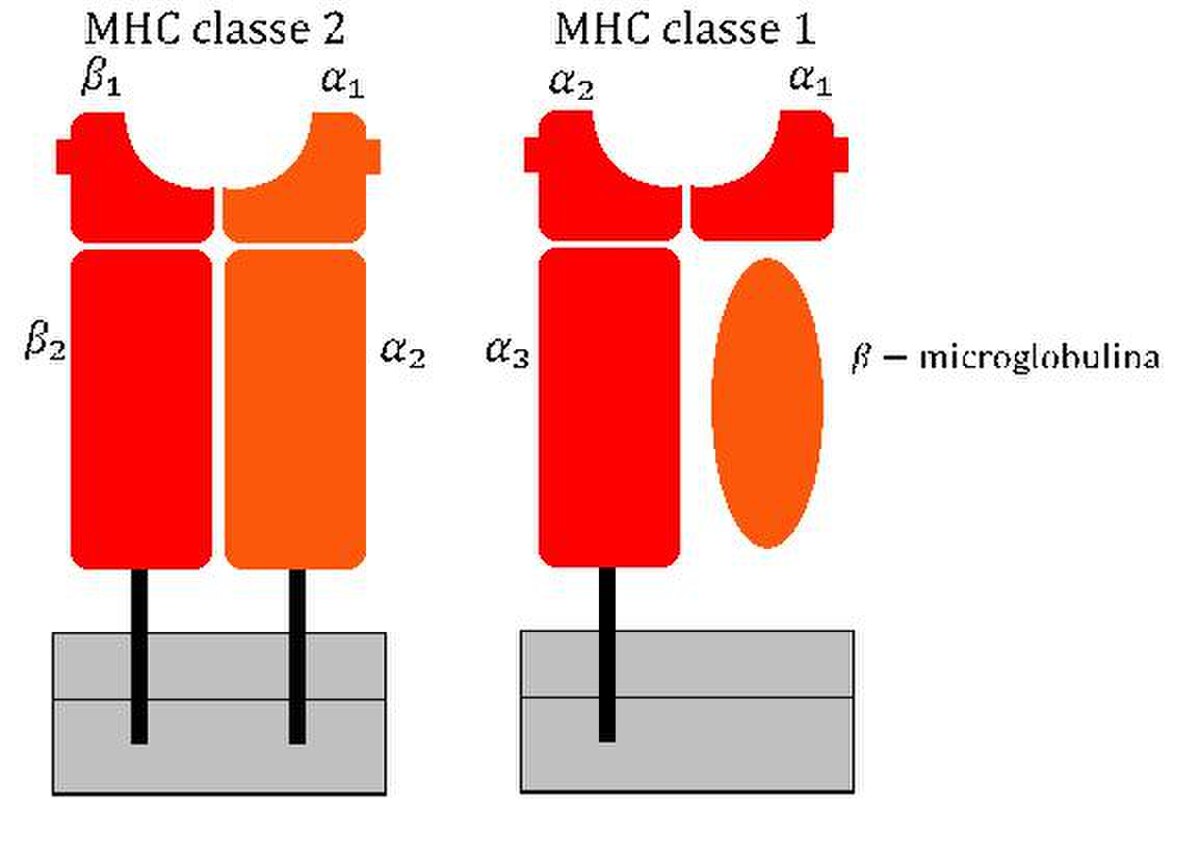
This schematic shows the structural differences between MHC class I and class II molecules used to present protein fragments on cell surfaces. BQmUB2011048, CC BY-SA 3.0, via Wikimedia Commons
When a healthy cell displays its own normal proteins, the immune system leaves it alone. If an intracellular pathogen such as a virus, Mycoplasma, or Rickettsia invades, MHC class I molecules display foreign pathogen fragments instead. Cytotoxic killer T cells use CD8 receptors and T-cell receptors to dock with the MHC molecule. If the receptor matches the displayed peptide, the killer T cell triggers the infected cell to destroy itself through apoptosis.
Why MHC genes vary between individuals
MHC genes are among the most diverse in the entire genome. In humans, the MHC region sits on chromosome 6, containing 224 genes across 3.6 million base pairs, and is commonly called the HLA complex.
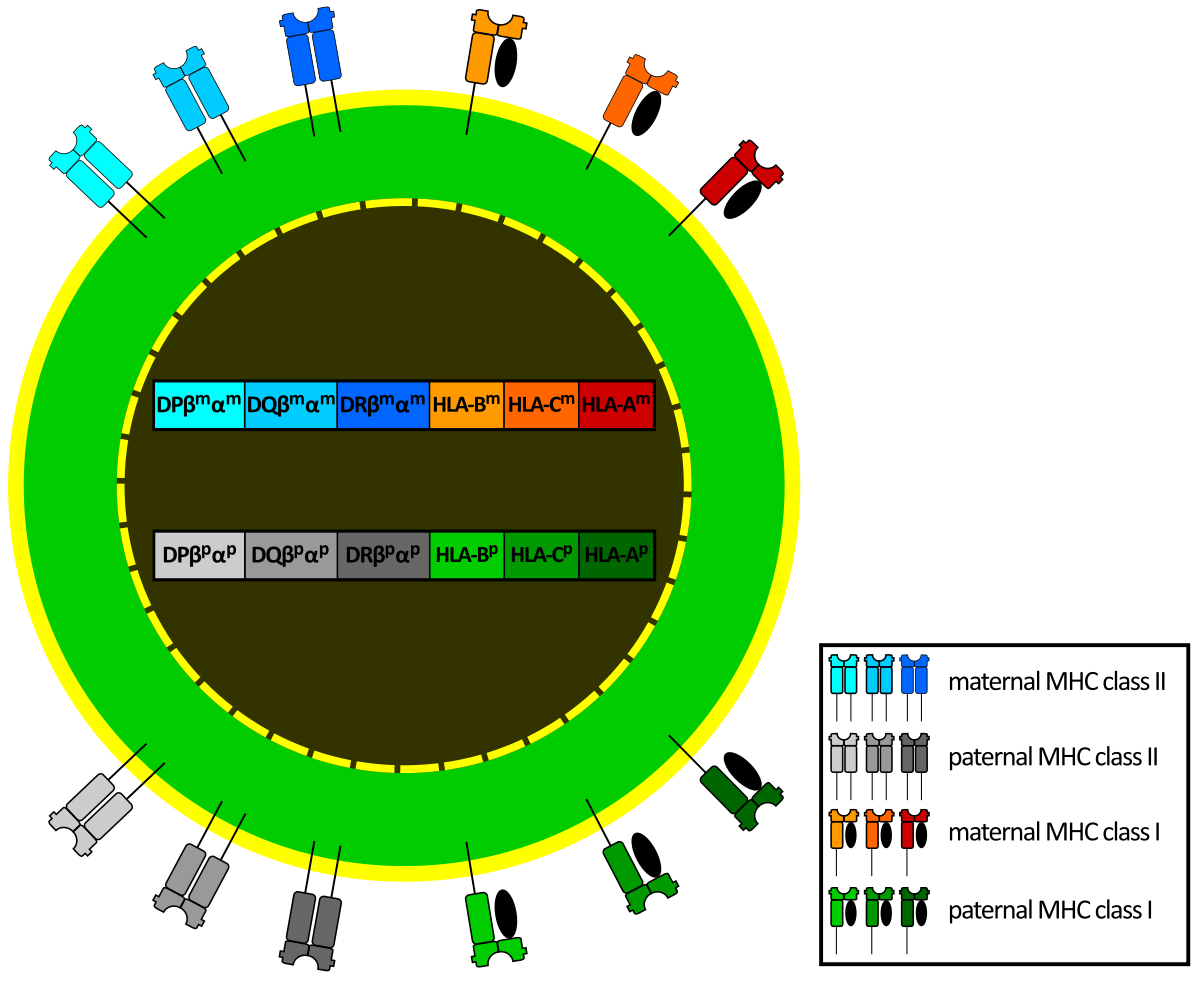
This diagram illustrates codominant expression, where alleles inherited from both parents are simultaneously expressed on cells. derivative work: Zionlion77 (talk) MHC_Class_1.svg: User atropos235 on en.wikipedia MHC_Class_2.svg: User atropos235 on , CC BY-SA 3.0, via Wikimedia Commons
Diversity comes from three genetic mechanisms: the system is polygenic with multiple interacting genes, expression is codominant using alleles from both parents, and the genes are highly polymorphic across the population. Over 19,000 distinct class I HLA alleles have been recorded in humans. This extreme variation is why finding matching donor organs for transplantation is difficult, and it even influences mate choice in mice.
Test yourself
What does the Major Histocompatibility Complex primarily signal to T cells?
The internal health of the cell. MHC displays protein fragments from inside the cell to reveal its current state. It acts as an ID to spot hijacked or cancerous cells, not just to label tissue types.
Play the lesson in edgi and the card is yours. It lands on your Map next to the ideas it connects to, and turns from matte to foil to gold as you learn more around it.
MHC is the general name for this gene complex across all jawed vertebrates. HLA, which stands for human leukocyte antigen, is specifically the human version of the MHC system.
Which cells in the body express MHC class I molecules?
MHC class I molecules are present on all nucleated cells and platelets. Red blood cells lack nuclei and do not express them.
Who discovered the Major Histocompatibility Complex?
Peter Gorer made the first descriptions in 1936, and George Snell identified the MHC locus in mice by breeding strains with differing tissue compatibility. Snell shared the 1980 Nobel Prize in Physiology or Medicine with Jean Dausset and Baruj Benacerraf for their work on these cell surface structures.