Portal veinThe vein that bypasses the heart
The portal vein is a large blood vessel in the abdomen that carries blood from the digestive organs and spleen straight to the liver. Unlike standard veins that return blood directly to the heart, it routes blood through a second capillary bed in the liver so nutrients can be absorbed and toxins filtered before entering general circulation. It provides the vast majority of the liver's total blood supply.

By the edgi team We find the most surprising true thing about an idea and build a 60-second lesson around it.
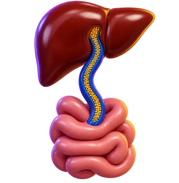
How the hepatic portal system works
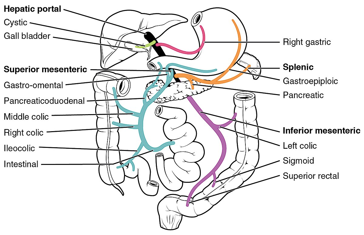
The portal vein is not a true vein because it does not empty directly into the heart. Instead, it forms a specialized bridge between two capillary networks, transporting blood from the stomach, intestines, pancreas, and spleen directly into the capillary-like sinusoids of the liver.
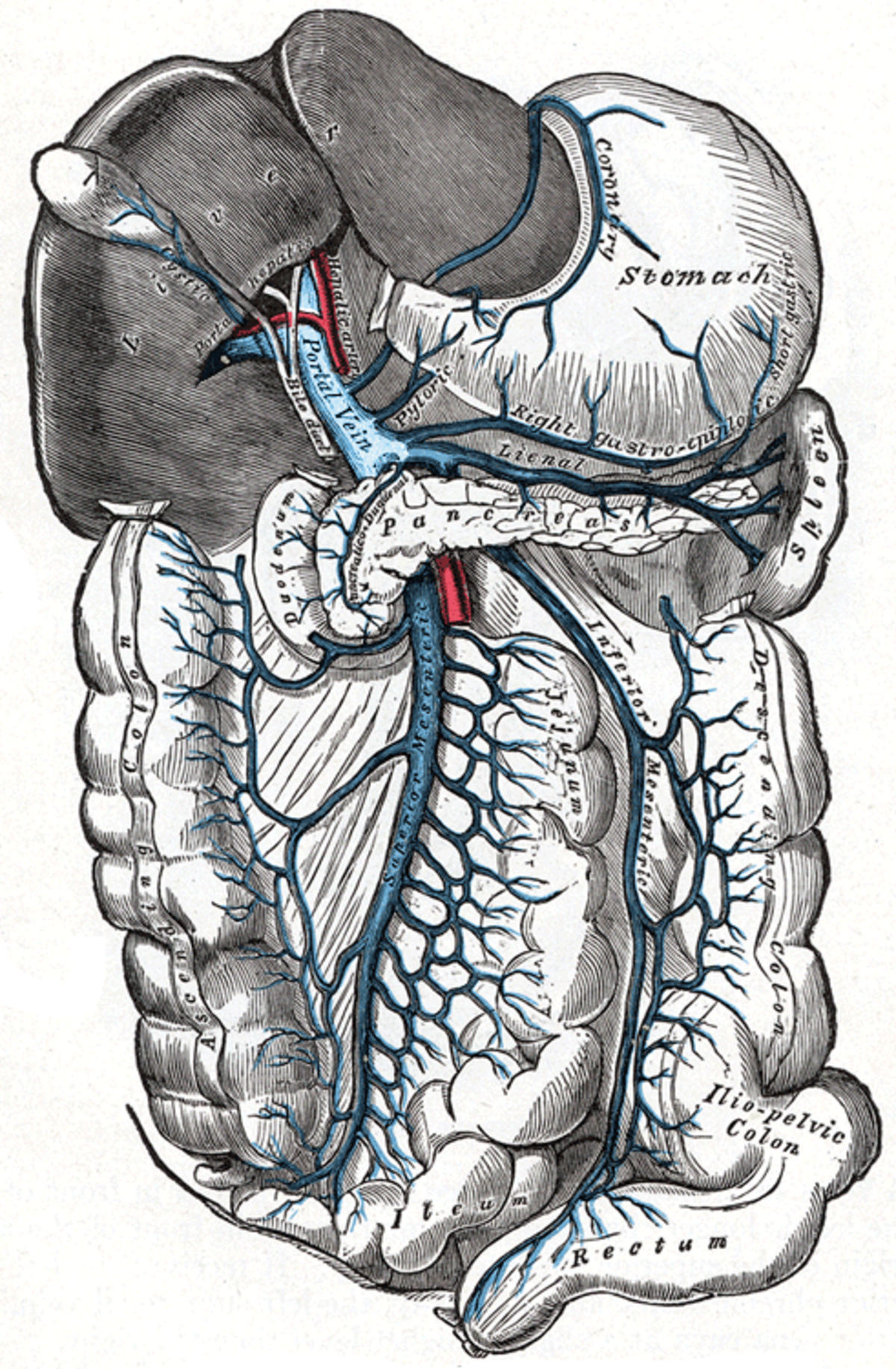
This detour serves two purposes. It ensures that the liver absorbs nutrients right after digestion, and it allows liver cells, called hepatocytes, to neutralize ingested toxins before blood reaches the rest of the body. Blood leaves these liver sinusoids through the hepatic veins, which then travel to the heart.
Together with the hepatic arteries, the portal vein forms a dual blood supply for the liver. The portal vein supplies 75% to 90% of the liver's blood flow and 70% of its oxygen. Because of this redundant blood supply, the liver suffers far less from vascular disease than other major internal organs.
Anatomy and branches
Measuring roughly 8 centimeters (3 inches) long in adults, the portal vein originates behind the neck of the pancreas in the upper right quadrant of the abdomen. In most people, it forms at the splenic-mesenteric confluence where the splenic vein and superior mesenteric vein meet.
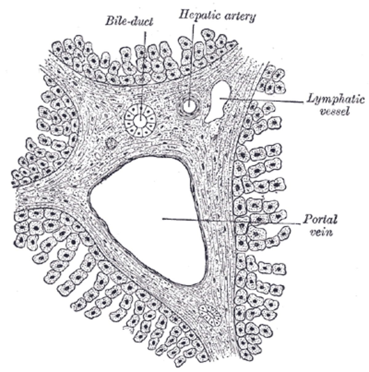
Other tributaries join along its path, including the left and right gastric veins, cystic veins, and pancreatic veins. Just before reaching the liver, the main trunk divides into right and left branches. These split into microscopic portal venules, which pair with hepatic arterioles to build the portal triads that feed the liver's tissue.
Small accessory hepatic portal veins also exist. These vessels, including the paraumbilical veins and veins from the gallbladder wall, drain straight into the liver without joining the main portal trunk.
Associated medical conditions
Cirrhosis often leads to portal hypertension, a dangerous rise in blood pressure inside the portal vein. Doctors diagnose this on Doppler ultrasound when the vessel dilates beyond 13 to 15 millimeters or when blood flow velocity drops below 16 centimeters per second.
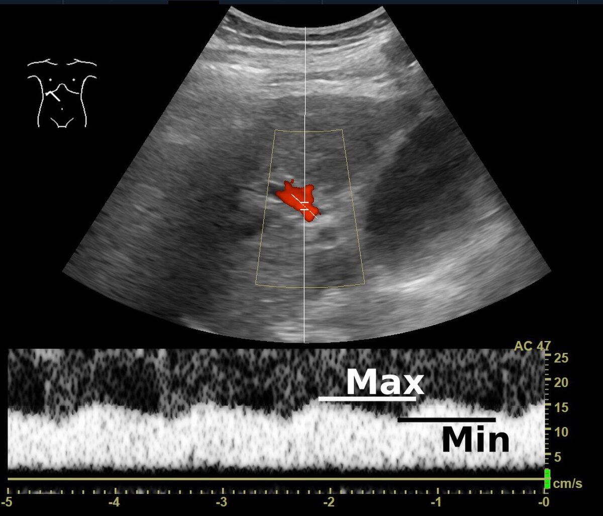
High pressure forces blood backward into connections with the systemic venous system, causing complications such as ascites, spider nevi, caput medusae, and enlarged esophageal varices that can rupture. Increased portal pulsatility can also indicate cirrhosis or high right atrial pressure from right heart failure.
In abdominal obesity, the portal vein carries fats and inflammatory substances from visceral fat into the liver, causing insulin resistance and metabolic dysfunction–associated steatotic liver disease. Other rare disorders include pylephlebitis, which is an infection of the vein usually triggered by conditions like diverticulitis, and portal venous gas, often caused by intestinal ischemia.

Collectible card
Claim the Portal vein card
Play the lesson in edgi and the card is yours. It lands on your Map next to the ideas it connects to, and turns from matte to foil to gold as you learn more around it.
Questions people ask
What other portal venous systems exist in the human body?
Humans have only two portal venous systems: the hepatic portal system and the hypophyseal portal system, which connects the hypothalamus to the pituitary gland. Some reptiles also possess a third type, called a renal portal system, which is absent in mammals.
What is pylephlebitis?
Pylephlebitis is an infection and inflammation of the portal vein. It typically develops as a complication of an intra-abdominal infection, such as diverticulitis.
What causes gas to appear in the portal vein?
Hepatic portal venous gas is a rare radiological sign where gas bubbles enter the portal circulation. It is most commonly caused by intestinal ischemia, though it is also linked to colon cancer.
Part of the Set · 9 cards
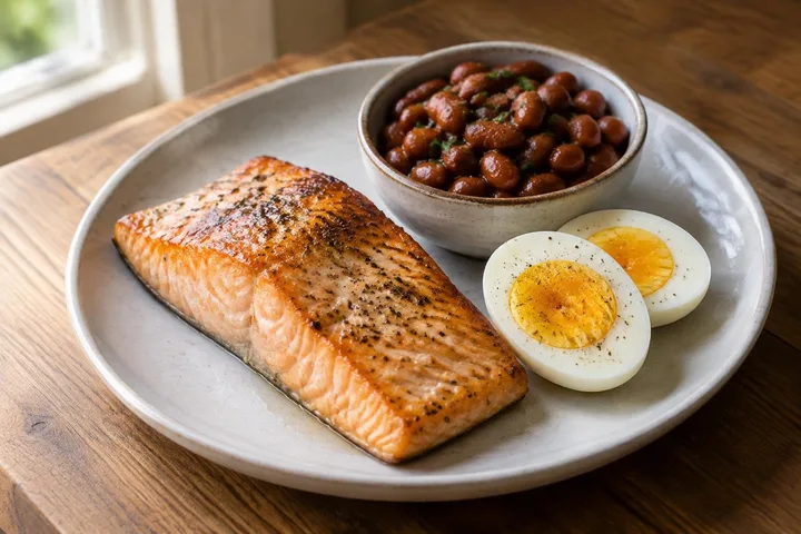
Your Body Never Sees Food
You ate a sandwich. Nothing that reached your blood had ever been a sandwich.
Learn the whole SetWhere this leads
- Blood Sugar LevelsBlood sugar measures circulating glucose, maintained at about one teaspoon in blood to feed the brain and tissues without causing toxic damage.
- Essential amino acidHumans cannot synthesize nine of the amino acids used to build proteins, meaning they must be supplied entirely by the foods in your diet.
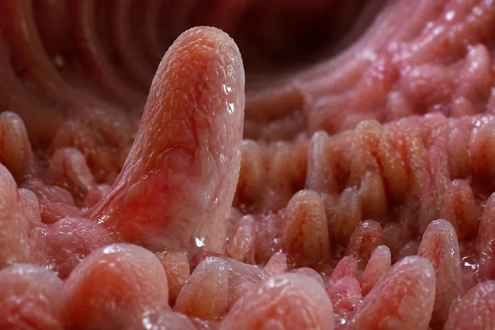
- Intestinal villusAn intestinal villus is a tiny, finger-like projection in the small intestine that increases surface area to absorb nutrients into the bloodstream.
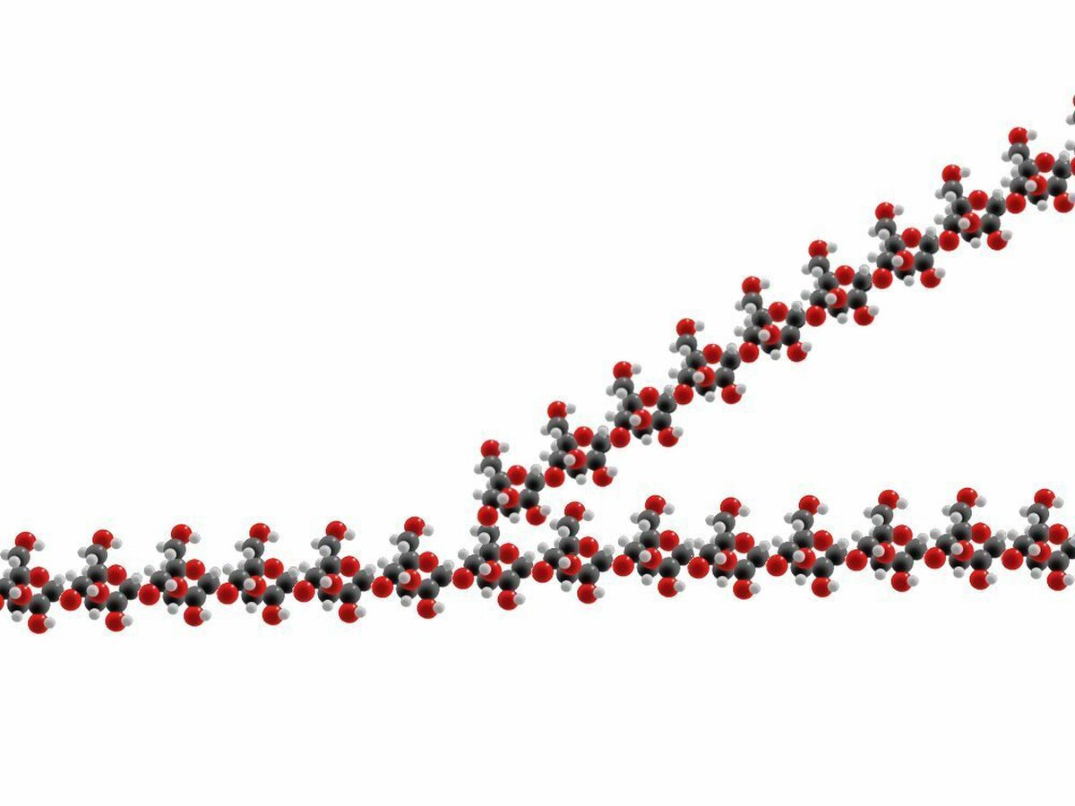
- GlycogenGlycogen is a dense, branched glucose chain stored in liver and muscle cells to supply rapid energy when blood sugar drops.
