Intestinal villusHow tiny folds create huge surface area
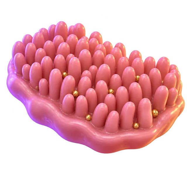
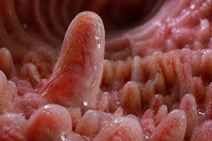
An intestinal villus is a tiny, finger-like projection lining the inside wall of the small intestine. Millions of them work together to expand the gut's surface area to roughly the size of a tennis court. This massive surface lets your digestive system absorb nutrients quickly through short-distance diffusion.
By the edgi team We find the most surprising true thing about an idea and build a 60-second lesson around it.
If you flattened out the inside of your small intestine, it would only look like a smooth cylinder about the size of a garden hose. Instead, nature packed it with wrinkles, folds, and millions of tiny finger-like projections called villi.
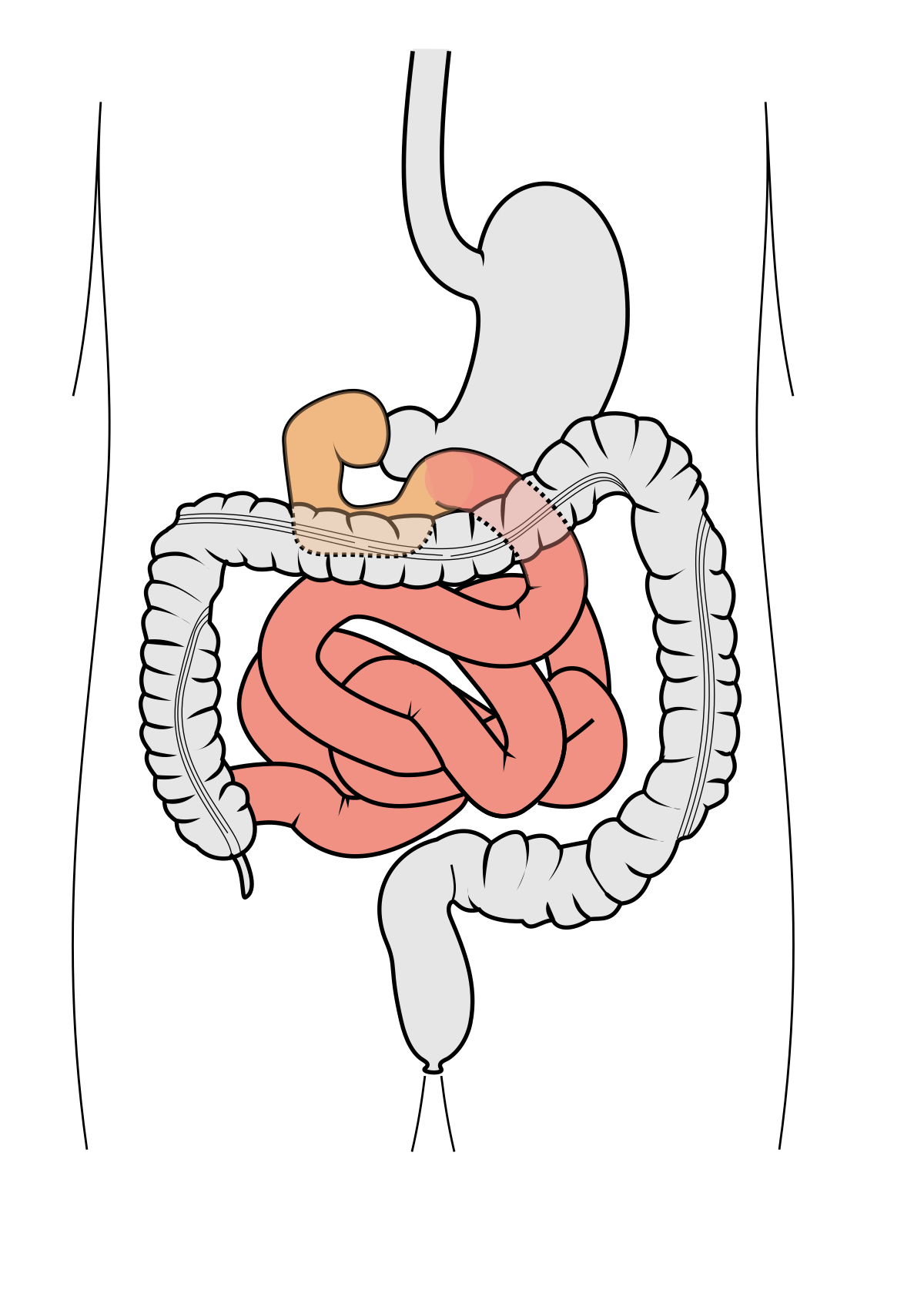
Diagram of the human digestive system focusing on the small intestine, highlighted in red, within the abdominal cavity. Olek Remesz (wiki-pl: Orem, commons: Orem), CC BY-SA 2.5, via Wikimedia Commons
Those microscopic folds expand your gut's inner surface area to roughly the size of a tennis court.
How it works
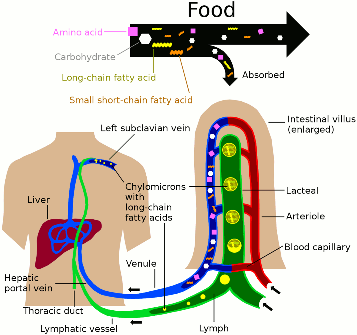
Each villus is only about a millimeter long, but it acts as a dedicated absorption tower for passing nutrients. Inside every villus sits a network of tiny blood vessels ready to catch sugars and amino acids.
Diagram illustrating nutrient absorption into the blood and lymph systems, showing food breakdown into Amino acid, Carbohydrate, Long-chain fatty acid, and Small short-chain fatty acid. Keministi, CC0, via Wikimedia Commons
Right alongside them are lymphatic vessels that absorb dietary fats directly into your circulation.
The brush border
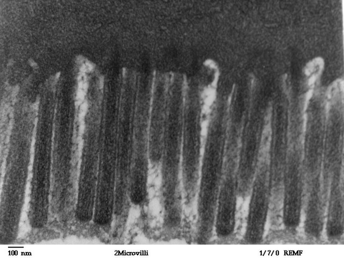
Zoom in even closer on a single villus, and you will find its outer cells are covered in microscopic hairs called microvilli. This dense layer is known as the brush border, and it pushes the available absorptive surface area even further.
A Transmission Electron Micrograph (TEM) shows the microvilli lining the human jejunum, appearing as numerous finger-like projections. Louisa Howard, Katherine Connollly, Public domain, via Wikimedia Commons
Without this microscopic architecture, your body could never pull enough nutrients out of your meals to keep you alive.
How villi absorb nutrients
Each intestinal villus is between 0.5 and 1.6 millimeters long, with walls only one cell thick. That ultra-thin barrier creates an extremely short diffusion path, letting digested food pass rapidly out of the intestinal cavity.
Inside each villus, specialized transport routes handle specific nutrients. Capillaries absorb simple sugars and amino acids directly into the bloodstream, while lymphatic capillaries called lacteals collect fats packaged as chylomicrons. Enterocytes, the main epithelial cells lining the villi, also carry digestive enzymes on their surfaces to break down food on contact.
The brush border and surface area multiplication
Villi expand the absorptive surface of the small intestine by roughly 40-fold on their own. The cells covering each villus are packed with thousands of microscopic hairs called microvilli, which are only about 1 micrometer long.
These microvilli form a dense layer known as the brush border. Together with the villi, this microscopic folding increases the intestine's total absorptive surface area around 600-fold, keeping nutrient uptake efficient as food moves past.
Test yourself
What structural role does the brush border play on an intestinal villus?
It pushes absorptive surface area further. The microvilli of the brush border create an even denser layer of microscopic hairs that further maximize the gut's surface area for absorption.
How do intestinal villi maximize nutrient uptake?
By dramatically expanding inner surface area. Intestinal villi act as microscopic folds that expand the gut lumen into a massive absorptive surface area, rather than acting primarily as enzyme secretors.
Why is the microscopic architecture of intestinal villi essential for human survival?
A smooth gut wall lacks sufficient area. Without the folds and villi turning a smooth garden hose into a tennis court of space, the body cannot absorb enough nutrients to stay alive.
Play the lesson in edgi and the card is yours. It lands on your Map next to the ideas it connects to, and turns from matte to foil to gold as you learn more around it.
Inflammation in the small intestine can flatten or completely destroy villi, a condition known as villous atrophy. This deterioration is often a feature of coeliac disease and drastically reduces the gut's ability to absorb nutrients.
Which cells make up an intestinal villus?
The epithelium covering each villus consists primarily of enterocytes and goblet cells. Enterocytes handle digestion and nutrient absorption, while goblet cells sit alongside them in the tissue layer.